The human gastrointestinal tract represents the most densely populated microbial ecosystem on the planet, harboring over (10^{13}) symbiotic microorganisms encoding millions of non-redundant genes. Rather than triggering devastating systemic inflammatory cascades, the mucosal immune system orchestrates an exquisite state of active immunological tolerance—harvesting metabolic benefits while maintaining an impenetrable barrier against microbial translocation.
1. The Biochemical Architecture of the Gut Mucosal Barrier
The intestinal epithelium is physically shielded from luminal microbes by a specialized biphasic mucus barrier produced by goblet cells:
- Outer Mucus Layer: Loosely organized, serving as the metabolic habitat and carbon source for commensal taxa (e.g., Akkermansia muciniphila, Bacteroides thetaiotaomicron).
- Inner Mucus Layer: Densely cross-linked, sterile gel of MUC2 mucin polymers anchored firmly to the epithelial apical surface, saturated with antimicrobial peptides (AMPs: (alpha)-defensins / cryptdins secreted by Paneth cells, RegIII(gamma), and lysozyme).
2. Secretory IgA (sIgA) Transcytosis and Immune Exclusion
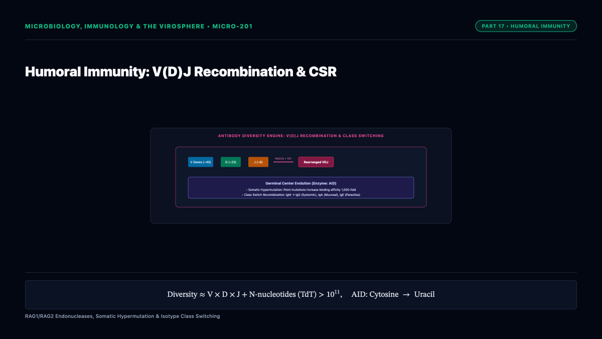
The primary humoral guardian of mucosal surfaces is dimeric Secretory Immunoglobulin A (sIgA). Mucosal plasma cells in the lamina propria produce dimeric IgA covalently joined by a joining (J) chain. The basal surface of enterocytes expresses the Polymeric Immunoglobulin Receptor (pIgR):
[
ext{Dimeric IgA} + ext{pIgR} xrightarrow{ ext{Endocytosis}} ext{Transcytosis to Apical Lumen} xrightarrow{ ext{Cleavage}} ext{sIgA} + ext{Secretory Component (SC)}
]
The cleaved extracellular domain of pIgR remains attached as the Secretory Component, shielding sIgA against harsh luminal proteolytic degradation. sIgA executes immune exclusion: cross-linking bacteria into agglutinated clumps, neutralizing toxins, and preventing microbial adhesion to epithelial apical membranes without inducing destructive complement activation.
3. Microbial Metabolites: Short-Chain Fatty Acids (SCFAs) as Immune Modulators
Anaerobic fermentation of non-digestible dietary polysaccharides by commensal phyla (principally Bacillota and Bacteroidota) yields substantial millimolar concentrations of Short-Chain Fatty Acids (SCFAs): acetate (( ext{C}_2)), propionate (( ext{C}_3)), and butyrate (( ext{C}_4)).
SCFAs act as powerful signaling ligands via G-protein coupled receptors (GPR41 / FFA3, GPR43 / FFA2, GPR109A) and as epigenetic modulators via direct inhibition of Histone Deacetylases (HDACs):
- Butyrate: Serves as the primary metabolic energy substrate for colonic enterocytes, driving mitochondrial (beta)-oxidation to consume local oxygen, thereby preserving the profound hypoxia (( ext{pO}_2 < 1 ext{ mmHg})) essential for obligate anaerobes.
- Epigenetic Induction of Tregs: Butyrate and propionate penetrate lamina propria dendritic cells and T cells, inhibiting HDACs (HDAC1/3). This hyperacetylates the Foxp3 locus enhancer (CNS1), profoundly stimulating the differentiation of anti-inflammatory peripherally-derived Regulatory T cells (( ext{pTregs})) that secrete IL-10 to suppress aberrant mucosal inflammation.
4. Tolerogenic Dendritic Cells and Peyer’s Patches
Specialized mucosal sampling occurs across follicle-associated epithelium via Microfold (M) cells, which lack microvilli and actively transcytose intact luminal antigens directly into subepithelial domes containing dense networks of ( ext{CD103}^+) Dendritic Cells (DCs).
Bathed in epithelial-derived TGF-(beta), Retinoic Acid (RA), and Thymic Stromal Lymphopoietin (TSLP), mucosal ( ext{CD103}^+) DCs adopt a stably tolerogenic phenotype. Upon capturing harmless commensal antigens, they migrate to mesenteric lymph nodes (MLNs), presenting antigen while synthesizing retinoic acid via retinal dehydrogenases (RALDH). Retinoic acid imprints gut-homing integrins ((alpha_4beta_7)) and chemokine receptors (CCR9) onto developing naïve T cells, directing them back to the gut lamina propria as immunosuppressive Tregs.