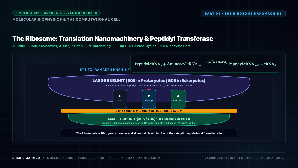
The ribosome is the universal molecular factory of biological translation—a massive $2.5text{–}4.5 text{ MDa}$ ribonucleoprotein machine that reads genetic instructions encoded in mRNA and polymerizes amino acids into proteins at velocities of $15text{–}20$ peptide bonds per second. Crucially, high-resolution atomic crystallography reveals that the catalytic Peptidyl Transferase Center (PTC) is entirely devoid of protein residues: the ribosome is a ribozyme.
1. Subunit Architecture & Dynamic Functional Sites
Across all kingdoms of life, the ribosome consists of two asymmetric subunits that associate during initiation and dissociate following termination:
- Bacterial 70S Ribosome:
- Small Subunit (30S): Composed of 16S ribosomal RNA ($sim 1,540 text{ nt}$) and 21 ribosomal proteins (S1–S21). Houses the Decoding Center where codon-anticodon fidelity is surveyed.
- Large Subunit (50S): Composed of 23S rRNA ($sim 2,900 text{ nt}$), 5S rRNA ($120 text{ nt}$), and 33 proteins (L1–L36). Houses the Peptidyl Transferase Center (PTC) and the expanding polypeptide exit tunnel.
- Eukaryotic 80S Ribosome:
- Small Subunit (40S, 18S rRNA) and Large Subunit (60S, 28S, 5.8S, 5S rRNAs) exhibiting conserved core architectures embellished with expanded eukaryotic rRNA insertion segments.
The inter-subunit interface defines three critical tRNA binding pockets:
- A-site (Aminoacyl-tRNA): Accommodates the incoming aminoacylated tRNA bearing the cognate anticodon triplet.
- P-site (Peptidyl-tRNA): Binds the tRNA carrying the growing nascent polypeptide chain.
- E-site (Exit): Holds the deacylated tRNA prior to its thermal release into the cytoplasm.
2. Hopfield-Ninio Kinetic Proofreading Mechanism
The standard free-energy difference between a cognate codon-anticodon pairing ($3$ base pairs) and a near-cognate mismatch ($2$ base pairs) is modest:
[
Delta Delta G^circ = Delta G^circ_{text{non-cognate}} – Delta G^circ_{text{cognate}} approx 2text{–}4 ; k_B T
]
Under simple equilibrium thermodynamics, the expected error rate would be:
[
eta_{text{eq}} = e^{-Delta Delta G^circ / k_B T} approx 10^{-1} text{ to } 10^{-2}
]
An error rate of $1%$ would result in catastrophic translational failure, as an average $400$-residue protein would contain multiple erroneous amino acids. In reality, the observed ribosomal error rate is extraordinarily low: $eta approx 10^{-4}$.
This fidelity is accomplished via the Hopfield-Ninio Kinetic Proofreading Mechanism, driven by the irreversible GTPase hydrolysis cycle of Elongation Factor Tu (EF-Tu):
[
text{Ribosome} + text{Ternary Complex} underset{k_{-1}}{overset{k_1}{rightleftharpoons}} C_{text{initial}} xrightarrow{k_2 text{ (GTP Hydrolysis)}} C_{text{activated}} xrightarrow{k_3 text{ (Accommodation)}} text{Peptide Bond}
]
[
C_{text{activated}} xrightarrow{w text{ (Dissociation / Proofreading)}} text{Reject}
]
Because the kinetic complex passes through an irreversible chemical state (GTP hydrolysis to GDP $+; text{P}_i$), the substrate is subjected to two independent discriminant kinetic screening barriers. The composite error rate achieves a squared fidelity ratio:
[
eta_{text{proofreading}} approx left( frac{k_{-1}^{text{near}}}{k_{-1}^{text{cognate}}} right) cdot left( frac{w^{text{near}}}{w^{text{cognate}}} right) approx e^{-2 Delta Delta G^circ / k_B T} approx 10^{-4}
]
3. The Catalytic Peptidyl Transferase Center: An Entropic Trap
When the 2.4 Å crystal structure of the large ribosomal subunit was solved by Venkatraman Ramakrishnan, Thomas Steitz, and Ada Yonath, it revealed that no protein side-chain approaches closer than $18 text{ AA}$ to the active site. The synthesis of peptide bonds is catalyzed entirely by conserved RNA nucleotides (specifically A2451 and C2063 in 23S rRNA numbering).
Detailed biophysical and quantum mechanical simulations demonstrate that the ribosome does not employ general acid-base catalysis. Instead, the PTC functions as an Entropic Catalytic Trap:
- It freezes the conformational mobility of the incoming amino acid’s nucleophilic $alpha$-amino group and the ester carbonyl of the peptidyl-tRNA, pre-orienting them into an ideal transition-state geometry.
- It shields the reactive centers from water, precluding non-productive ester hydrolysis.
- The ribosome accelerates peptide bond formation by reducing the activation entropy $Delta S^ddagger$ by a factor of nearly $10^7$, with minimal contribution from activation enthalpy $Delta H^ddagger$.
4. Translocation Dynamics & The EF-G Molecular Ratchet
Following peptide bond synthesis, the ribosome finds itself in a hybrid state ($text{A/P}$ and $text{P/E}$). To prepare for the next translation cycle, the mRNA and tRNAs must advance precisely one triplet ($0.34 text{ nm}$) relative to the ribosome.
This step is catalyzed by Elongation Factor G (EF-G), a molecular GTPase motor. Cryo-EM reconstructions reveal that the ribosome undergoes a spontaneous thermal ratcheting motion: the small subunit rotates $6^circtext{–}9^circ$ counterclockwise relative to the large subunit. EF-G binds to this ratcheted intermediate, hydrolyzing GTP to decouple ribosomal inter-subunit bridges and mechanically propel the tRNAs into the canonical P and E sites, completing a cycle of translation with sub-nanometer precision.