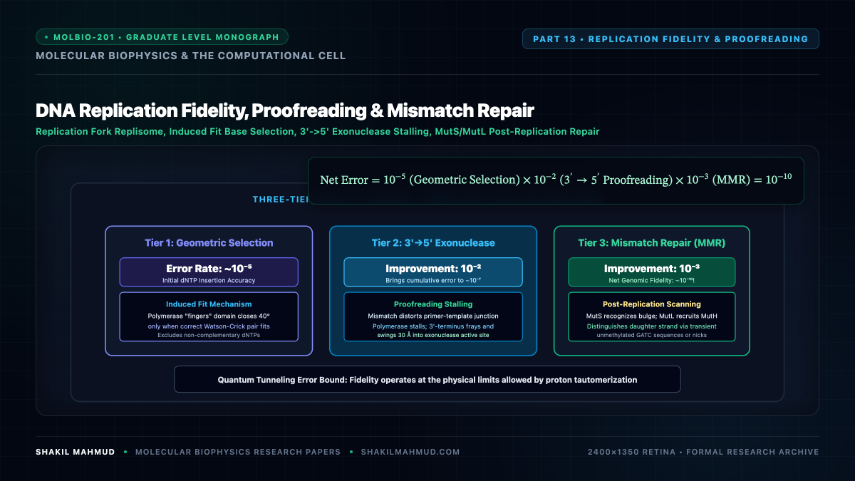
The human genome comprises $3.2 times 10^9$ base pairs. During somatic cell division, this massive molecular archive must be duplicated with near-miraculous fidelity: fewer than three mutations are introduced per complete replication cycle. Achieving an overall error rate of $sim 10^{-10}$ requires a multi-layered, thermodynamic-kinetic error-correction hierarchy operating across three sequential stages.
1. The Three-Tier Fidelity Cascade
The total error rate $epsilon_{text{total}}$ of DNA replication is the multiplicative product of three independent molecular screening filters:
[
epsilon_{text{total}} = epsilon_{text{selectivity}} times epsilon_{text{proofreading}} times epsilon_{text{MMR}} approx 10^{-5} times 10^{-2} times 10^{-3} approx 10^{-10}
]
- Active-Site Geometric Selectivity ($epsilon sim 10^{-4}text{–}10^{-5}$): Replicative DNA polymerases ($text{Pol } delta$ and $text{Pol } epsilon$ in eukaryotes, $text{Pol III}$ in prokaryotes) possess a rigid catalytic pocket shaped precisely to fit the Watson-Crick base pair geometry ($10.85 text{ AA}$ $text{C}1′-text{C}1’$ distance). Non-canonical pairs (purine-purine or pyrimidine-pyrimidine) cause steric clash or misalignment of the incoming $alpha$-phosphate relative to the catalytic $text{Mg}^{2+}$ ions, reducing the phosphodiester polymerization rate $k_{text{pol}}$ by $10^5$-fold.
- Exonucleolytic Proofreading ($epsilon sim 10^{-2}$): If a mismatched base is erroneously incorporated, terminal base-pairing disruption stalls the polymerase. The primer strand melts and partitions into the distinct $3′ to 5’$ exonuclease active site located $30text{–}40 text{ AA}$ away, where the incorrect nucleotide is catalytically excised.
- Post-Replicative Mismatch Repair ($epsilon sim 10^{-2}text{–}10^{-3}$): The MutS/MutL complex scans the nascent daughter duplex, recognizing distortions in the helical contour and initiating excision of the error-containing nascent strand.
2. Kinetic Modeling of Exonucleolytic Switching
The competition between forward elongation and exonuclease editing is modeled as a branched kinetic network:
[
E cdot D_n xrightarrow{k_{text{pol}}} E cdot D_{n+1} quad text{(Polymerization)}
]
[
E cdot D_n underset{k_{text{switch-back}}}{overset{k_{text{switch}}}{rightleftharpoons}} E_{text{exo}} cdot D_n xrightarrow{k_{text{exo}}} E_{text{exo}} cdot D_{n-1} quad text{(Proofreading)}
]
For a correctly matched terminus, $k_{text{pol}} gg k_{text{switch}}$, ensuring processive synthesis at speeds up to $1,000 text{ nt/s}$. For an incorrect mismatch terminus, base unpairing accelerates the strand-melting rate and structural switching to the exonuclease active site by $10^3$-fold ($k_{text{switch}} gg k_{text{pol}}$), routing the mispaired strand toward excision.
3. Post-Replication Mismatch Repair: Strand Discrimination Biophysics
To repair an erroneous base, the cell must solve a profound symmetry puzzle: how does the repair machinery discriminate between the parental template strand (which contains the correct historical sequence) and the nascent daughter strand (which harbors the error)?
- Prokaryotic Methylation-Directed Discrimination: In E. coli, the enzyme Dam methylase methylates adenine bases at $5’text{-GATC-3′}$ sequences. Immediately following replication, DNA is transiently hemimethylated. The endonuclease MutH selectively binds unmethylated daughter strands, nicking the newly synthesized strand for degradation by UvrD helicase and exonucleases.
- Eukaryotic PCNA-Directed Discrimination: Eukaryotes lack DNA adenine methylation. Instead, strand discrimination is physically guided by the asymmetric loading of the proliferating cell nuclear antigen (PCNA) sliding clamp and the pre-existing nicks between Okazaki fragments on the lagging strand, or transient ribonucleotide excision nicks on the leading strand. MutS$alpha$ (MSH2-MSH6) and MutL$alpha$ (MLH1-PMS2) assemble at the mismatch, using PCNA orientation to direct the latent endonuclease activity of PMS2 exclusively into the nascent daughter strand.