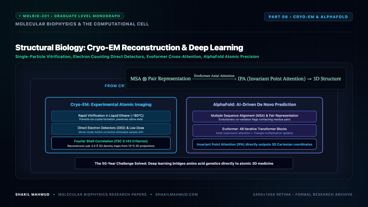
The structural determination of biological macromolecules has undergone two historic revolutions over the past decade: the experimental Resolution Revolution driven by Single-Particle Cryogenic Electron Microscopy (Cryo-EM), and the computational breakthrough of deep-learning-based structure prediction embodied by AlphaFold.
1. Single-Particle Cryo-EM & Contrast Transfer Function (CTF)
In cryogenic electron microscopy, biological samples in thin aqueous films are rapidly plunge-frozen into liquid ethane cooled to near liquid nitrogen temperatures ($approx -180^circ text{C}$ at cooling rates $> 10^5 text{ K/s}$). This vitrification process bypasses crystalline ice nucleation, trapping water and macromolecules in amorphous glass that preserves physiological native architectures.
When high-energy electrons ($200text{–}300 text{ keV}$, relativistic wavelength $lambda approx 0.02text{–}0.025 text{ AA}$) pass through the vitrified specimen, the image formed on direct electron detectors represents a 2D projection of the specimen’s electrostatic potential $phi(mathbf{r})$. In Fourier space, the recorded image intensity $I(mathbf{k})$ is modulated by the microscope’s Contrast Transfer Function (CTF):
[
text{CTF}(mathbf{k}) = -A(mathbf{k}) sinleft( pi lambda Delta z |mathbf{k}|^2 + frac{pi}{2} C_s lambda^3 |mathbf{k}|^4 right)
]
Where:
- $Delta z$ is the objective lens defocus distance.
- $C_s$ is the spherical aberration coefficient of the electromagnetic objective lens.
- $A(mathbf{k})$ is the spatial coherence envelope function decaying at high spatial frequencies.
Because the $sin(cdot)$ term oscillates between positive and negative values with multiple zero-crossings, accurate 3D volume reconstruction strictly requires computational phase-flipping and CTF deconvolution across hundreds of thousands of noisy single-particle micrographs.
2. 3D Fourier Slice Theorem & Resolution Assessment
The mathematical foundation of 3D volume reconstruction from 2D micrographs is the Projection-Slice Theorem (Central Slice Theorem): the 2D Fourier transform $mathcal{F}_{2D}$ of a projection of a 3D electrostatic density $rho(x, y, z)$ along projection vector $hat{mathbf{n}}$ is mathematically identical to a 2D central slice through the 3D Fourier transform $mathcal{F}_{3D}[rho]$ perpendicular to $hat{mathbf{n}}$:
[
mathcal{F}_{2D}[P_{hat{mathbf{n}}}rho](k_x, k_y) = mathcal{F}_{3D}[rho](k_x, k_y, 0)
]
To determine the true atomic resolution of the reconstructed 3D map, the dataset of single particles is randomly partitioned into two independent halves (gold-standard split). The Fourier Shell Correlation (FSC) between the two independent 3D reconstructions $rho_1$ and $rho_2$ is computed over concentric spherical shells of radius $k$ in Fourier space:
[
text{FSC}(k) = frac{sum_{|mathbf{k}| = k} F_1(mathbf{k}) cdot F_2^*(mathbf{k})}{sqrt{sum |F_1(mathbf{k})|^2 cdot sum |F_2(mathbf{k})|^2}}
]
By international structural biology convention, the nominal map resolution is defined at the frequency where $text{FSC}(k) = 0.143$, reflecting an informational signal-to-noise ratio sufficient to model atomic coordinate positions with sub-angstrom precision.
3. AlphaFold 2 Architecture: The Evoformer & Invariant Point Attention
In CASP14 (2020), DeepMind’s AlphaFold 2 solved the 50-year grand challenge of predicting 3D protein structures from primary amino acid sequence with experimental atomic accuracy ($text{RMSD} lesssim 1.5 text{ AA}$).
AlphaFold 2 replaced hand-crafted physical force fields with a differentiable end-to-end neural network architecture featuring two primary innovations:
- The Evoformer: A specialized transformer block that operates simultaneously on two representations:
- Multiple Sequence Alignment (MSA) Representation: Encoding evolutionary co-variation across millions of homologous genomes ($N_{text{seq}} times N_{text{res}}$). Correlated mutations between residue positions $i$ and $j$ reveal physical spatial proximity.
- Pair Representation: Encoding spatial geometric relationships ($N_{text{res}} times N_{text{res}}$) between all residue pairs.
Information flows bidirectionally between the MSA and Pair representations using row/column axial self-attention and outer-product triangular updates that enforce Euclidean geometric triangular inequalities:
[
d_{ik} le d_{ij} + d_{jk}
] - The Structure Module & Invariant Point Attention (IPA): Operates directly in 3D Euclidean space. It represents the protein backbone as a continuous chain of rigid geometric frames $T_i = (R_i, mathbf{t}_i) in text{SE}(3)$ (rotation matrix $R_i in text{SO}(3)$ and translation vector $mathbf{t}_i in mathbb{R}^3$). The IPA mechanism updates residue coordinates while maintaining strict equivariance under rigid-body spatial rotations and translations.
4. Confidence Metrics: pLDDT and PAE Matrices
AlphaFold provides explicit Bayesian uncertainty estimates for every predicted coordinate:
- pLDDT (Predicted Local Distance Difference Test): A per-residue score on a scale from $0$ to $100$:
[
text{pLDDT} > 90 implies text{High-accuracy atomic coordinates (comparable to } le 2.0text{AA crystal structures)}.
]
Regions with $text{pLDDT} < 50$ correlate strongly with naturally intrinsically disordered regions (IDRs). - Predicted Aligned Error (PAE): An $N times N$ matrix where entry $(i, j)$ estimates the expected error in the 3D position of residue $j$ when the predicted structure is aligned onto residue $i$. Low PAE between domains indicates confident relative rigid-body orientation.