DNA is far more than a passive archival tape of biological data. In the dense interior of the living cell, DNA behaves as an elastic rod governed by non-linear mechanics, differential geometry, and topology. Its helical geometry and topological supercoiling dictate gene accessibility, transcriptional elongation, and chromosomal segregation.
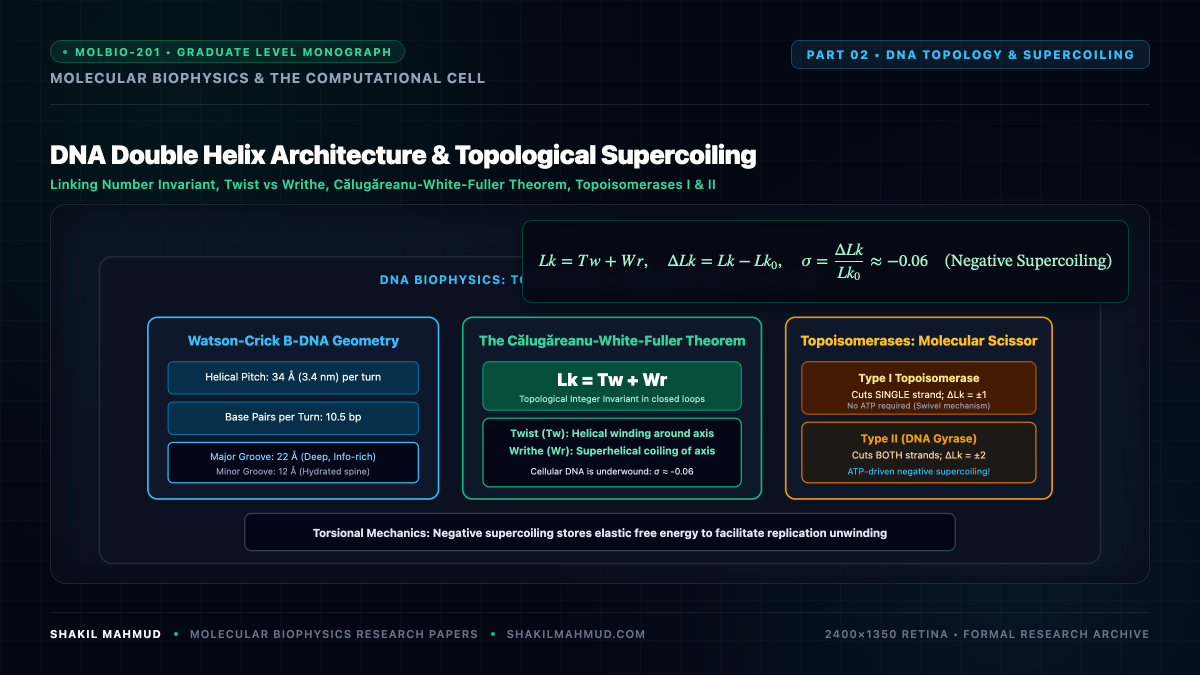
1. The Watson-Crick B-DNA Geometric Parameters
In standard aqueous physiological conditions (high water activity, physiological ionic strength $sim 150 text{ mM NaCl}$), duplex DNA crystallizes predominantly in the right-handed B-DNA conformation:
- Helical Diameter: $2.0 text{ nm}$ ($20 text{ AA}$).
- Helical Pitch: $3.4 text{ nm}$ per complete $360^circ$ rotation.
- Base Pairs per Turn: $sim 10.5 text{ bp}$ in liquid solution.
- Axial Rise per Base Pair: $h = 0.34 text{ nm}$ ($3.4 text{ AA}$).
- Rotational Angle per Base Pair: $Delta theta approx 34.3^circ$.
- Major Groove vs Minor Groove: Major groove width $sim 1.2 text{ nm}$ (exposing rich chemical patterns for sequence-specific transcription factor hydrogen bonding); Minor groove width $sim 0.6 text{ nm}$.
The stability of the double helix is not primarily driven by the horizontal hydrogen bonds between purines and pyrimidines ($2$ for $text{A=T}$, $3$ for $text{G}equivtext{C}$); rather, it is thermodynamically dominated by vertical base stacking interactions. Overlapping aromatic $pi$-electron orbitals between adjacent heterocyclic planar rings contribute negative free energy ($Delta G_{text{stacking}}^circ sim -1.0 text{ to } -3.5 text{ kcal/mol}$), reinforced by favorable entropic expulsion of structured hydration water shells into bulk solvent.
2. The Călugăreanu-White-Fuller Topological Invariant
When the two complementary single strands of a circular closed DNA molecule (such as a bacterial plasmid or topologically constrained eukaryotic chromatin loop) wrap around each other, their topological relationship becomes invariant under any smooth deformation that does not cleave the phosphodiester backbone.
This topological constraint is formalized by the Călugăreanu-White-Fuller Theorem:
[
Lk = Tw + Wr
]
Where:
- $Lk$ (Linking Number): An integer topological invariant representing the number of times one strand pierces the surface bounded by the other strand. It can only change if covalent chemical bonds are broken and reformed by topoisomerase enzymes.
- $Tw$ (Twist): A geometric measure of the helical rotation of the strands about the duplex central axis:
[
Tw = frac{1}{2pi} int_0^L left( mathbf{n} times frac{dmathbf{n}}{ds} right) cdot mathbf{t} , ds approx frac{N}{10.5}
]
where $N$ is the total number of base pairs. - $Wr$ (Writhe): A non-local geometric measure of the self-coiling and spatial contortion of the duplex axis in three-dimensional space, defined by the Gauss double line integral:
[
Wr = frac{1}{4pi} oint_{C} oint_{C} frac{(mathbf{r}_1 – mathbf{r}_2) cdot (dmathbf{r}_1 times dmathbf{r}_2)}{|mathbf{r}_1 – mathbf{r}_2|^3}
]
3. Supercoiling Density & Free Energy Storage
The relaxed linking number of a planar circular DNA molecule of $N$ base pairs is:
[
Lk_0 = frac{N}{10.5}
]
Any deviation from this relaxed state produces a linking difference $Delta Lk = Lk – Lk_0$. The non-dimensional superhelical density $sigma$ is defined as:
[
sigma = frac{Delta Lk}{Lk_0} = frac{Lk – Lk_0}{Lk_0}
]
In living bacterial cells such as Escherichia coli, the genome is maintained in a state of negative supercoiling, typically with:
[
sigma approx -0.06 quad (text{6% underwound})
]
The mechanical free energy $Delta G_{text{sc}}$ stored within a supercoiled DNA plasmid scales quadratically with the linking difference:
[
Delta G_{text{sc}} = frac{K_{text{topo}}}{N} (Delta Lk)^2 = 1050 , R T frac{(Delta Lk)^2}{N} = 10 cdot R T cdot N cdot sigma^2
]
This stored torsional elastic energy reduces the thermodynamic barrier required to melt the double helix ($Delta G_{text{unwinding}} < 0$), fundamentally enabling RNA polymerase open-complex formation during transcriptional initiation without requiring prohibitive ATP expenditure.
4. Topoisomerases: The Mechanical Maxwell Demons of the Genome
During DNA replication and transcriptional elongation, the moving replication fork or RNA polymerase motor acts as a torsional pump (described by the Liu-Wang Twin-Supercoiled-Domain model), accumulating positive supercoils ($+Wr$) ahead of the complex and negative supercoils ($-Wr$) behind it.
To relieve this catastrophic torsional strain, cells deploy Topoisomerases:
- Type I Topoisomerases (e.g., Topo I, Topo III): Introduce a transient single-strand break using a catalytic active-site tyrosine nucleophile, allowing the intact strand to rotate through the nick before religating, changing linking number in integer steps of $Delta Lk = pm 1$ without ATP hydrolysis.
- Type II Topoisomerases (e.g., DNA Gyrase, Topo IV, eukaryotic Topo II): Form a covalent double-strand gate, utilizing the energy of two ATP hydrolysis cycles to pass an intact double-stranded DNA segment ($T$-segment) completely through the gate ($G$-segment), altering the linking number in discrete topological quanta of:
[
Delta Lk = pm 2
]
Bacterial DNA gyrase is the unique molecular machine capable of actively pumping negative supercoils ($Delta Lk = -2$) into relaxed DNA against a thermodynamic gradient, keeping the bacterial chromosome primed for computational activity.