In 1944, Austrian physicist Erwin Schrödinger published a short, visionary book that would mesmerize an entire generation of physical scientists: What is Life? Schrödinger reasoned from quantum thermodynamics that living organisms must possess a microscopic “hereditary code-script” embedded within an aperiodic crystal—a long molecular polymer that resisted thermal fluctuations while packing astronomical quantities of genetic information into sub-cellular dimensions. The hunt for this molecular crystal became the holy grail of twentieth-century science.
By the early 1950s, biophysical experiments had eliminated proteins and unmasked deoxyribonucleic acid (DNA) as the transforming agent of genetic inheritance. Furthermore, Columbia University biochemist Erwin Chargaff (1905–2002) discovered a perplexing empirical constancy across all biological species—Chargaff’s Rules:
$$[text{Adenine}] = [text{Thymine}] quad text{and} quad [text{Guanine}] = [text{Cytosine}]$$
No matter the organism—whether bacteriophage, sea urchin, or human—the molar concentration of adenine always equaled thymine, and guanine always equaled cytosine. Yet no one understood the stereochemical geometry that dictated this quantitative equality.
Rosalind Franklin and Photo 51
The experimental resolution of the structure was achieved at King’s College London by the brilliant physical chemist and crystallographer Rosalind Elsie Franklin (1920–1958). Franklin was a master of high-resolution X-ray diffraction of non-crystalline paracrystalline fibers. By precisely controlling the relative humidity of delicate DNA fiber samples, Franklin discovered that DNA existed in two distinct structural states: the crystalline “A” form at lower hydration and the paracrystalline “B” form at high humidity ($>90%$), which represented DNA in its native physiological state.
In May 1952, after an exposure of over sixty hours in a hydrogen-filled camera, Franklin captured the legendary Photo 51. The photograph revealed an unmistakable dark, cross-shaped pattern of diffraction spots—the diagnostic signature of a helical structure—with missing layer lines on the fourth tier that proved the presence of two interlocking, helical strands offset by a specific minor and major groove.
The Watson-Crick Molecular Model (1953)
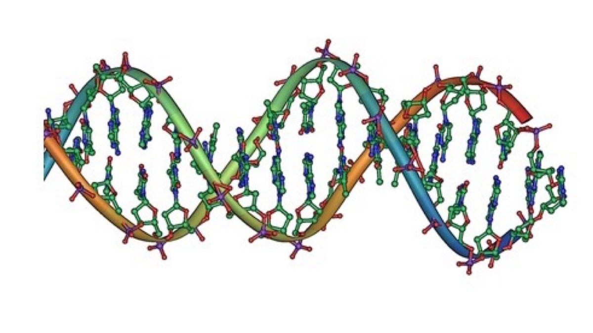
Armed with Franklin’s unpublished diffraction data and Chargaff’s ratios, twenty-four-year-old American biologist James Watson and British physicist Francis Crick at Cambridge’s Cavendish Laboratory constructed their iconic physical scale model in March 1953. The architectural breakthrough occurred when Watson realized that by orienting the purine and pyrimidine nitrogenous bases in their correct keto (rather than enol) tautomeric forms, complementary pairs formed perfectly identical geometrical dimensions held together by intermolecular hydrogen bonds:
- Adenine pairs with Thymine via two hydrogen bonds ($A = T$).
- Guanine pairs with Cytosine via three hydrogen bonds ($G equiv C$).
The two sugar-phosphate backbones ran in opposite, antiparallel directions ($5^prime rightarrow 3^prime$ and $3^prime rightarrow 5^prime$), wrapping around a central axis with a helical pitch of $34 text{ AA}$ (10 base pairs per complete turn). In their single-page announcement in Nature on April 25, 1953, Watson and Crick concluded with one of the most famous understatements in scientific history:
“It has not escaped our notice that the specific pairing we have postulated immediately suggests a possible copying mechanism for the genetic material.”
Because each base strictly paired with its complement, unzipping the double helix allowed each single strand to serve as an exact template for synthesizing a brand-new identical copy. The secret of life was not a mystical vitalist fluid; it was stereochemical hydrogen bonding governed by the laws of quantum chemistry.