Despite Oswald Avery’s rigorous demonstration in 1944 that DNA mediated bacterial transformation, skepticism persisted among elite biochemists. Many argued that pneumococcal transformation was a specialized bacterial anomaly, or that minute, undetectable traces of protein adhering to Avery’s DNA preparations were the true genetic messengers. To convert the scientific world, genetics required an experimental system of radical simplicity—an organism stripped of cellular complexity, reduced to the barest dualism of protein and nucleic acid. That ideal weapon was discovered in the alien, geometric architecture of the bacterial virus: the bacteriophage.
Under the intellectual leadership of Max Delbrück and Salvador Luria (founders of the famed “Phage Group”), researchers turned to Bacteriophage T2, an obligate parasite of the bacterium Escherichia coli. Under the newly invented transmission electron microscope, phage T2 looked like a lunar lander: an icosahedral protein head enclosing a dense macromolecular core, mounted upon a cylindrical contractile tail terminating in delicate tail fibers. Biochemically, phage T2 was remarkably pure: it consisted solely of approximately 50% protein and 50% DNA. When a phage attacked an E. coli cell, it attached to the bacterial wall, and twenty minutes later, the bacterium lysed open, releasing hundreds of identical progeny phages. But what entered the bacterial fortress to command this replication: the protein coat, or the DNA core?
The Radioactive Isotopes: 32P versus 35S
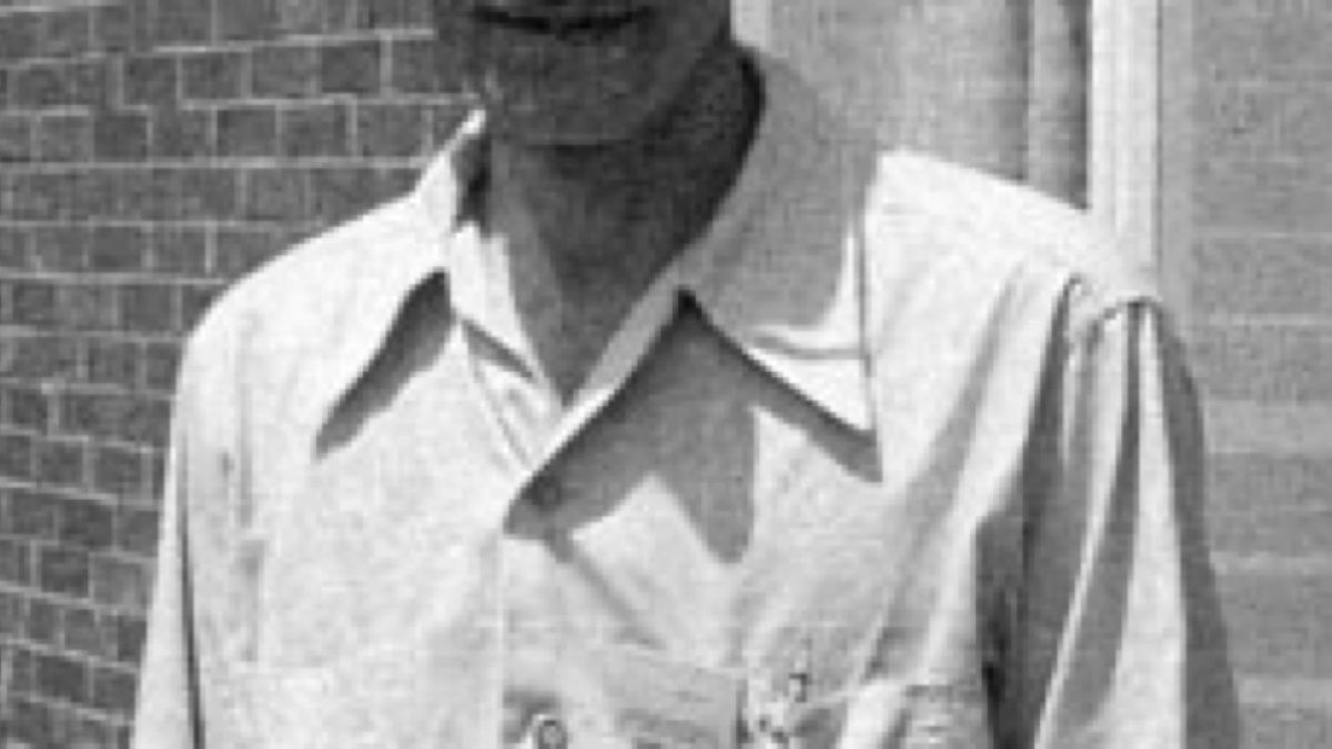
In the summer of 1952 at the Carnegie Institution of Washington’s Department of Genetics at Cold Spring Harbor, New York, geneticist Alfred Hershey (1908–1997) and his talented twenty-four-year-old laboratory assistant Martha Chase (1927–2003) designed an experiment of sublime physical elegance. Their strategy exploited a fundamental stoichiometric difference between proteins and nucleic acids:
- Phosphorus ($^{32}text{P}$): DNA contains enormous quantities of phosphorus within its repeating sugar-phosphate backbone, whereas proteins contain virtually zero phosphorus.
- Sulfur ($^{35}text{S}$): Proteins contain sulfur within the amino acids methionine and cysteine, whereas DNA contains zero sulfur atoms.
Hershey and Chase prepared two distinct batches of radioactive T2 bacteriophages:
- Batch 1 was cultured in E. coli growing in medium containing radioactive Phosphorus-32 ($^{32}text{P}$), producing phages with radioactively labeled DNA enclosed in non-radioactive protein coats.
- Batch 2 was cultured in medium containing radioactive Sulfur-35 ($^{35}text{S}$), producing phages with radioactively labeled protein coats enclosing non-radioactive DNA.
The Waring Blender and the Centrifuge
Hershey and Chase then permitted each radioactive phage batch to infect fresh, unlabelled E. coli cells in liquid suspension. Once the phages adsorbed to the bacterial membranes and initiated injection, Hershey and Chase faced a mechanical dilemma: how could they detach the empty viral ghost particles remaining on the bacterial exterior without lysing the infected cells?
Their solution became a legend of experimental physics: an ordinary electric Waring kitchen blender. By placing the infected bacterial suspension into the blender and agitating it at high speed for several minutes, the powerful shearing forces tore the viral coats off the cell walls without disrupting the heavier, intact bacterial bodies.
The agitated mixture was immediately transferred to a centrifuge. Centrifugation cleanly separated the components based on mass:
- The heavy, intact bacterial cells sedimented to the bottom of the tube, forming a solid pellet.
- The sheared, lightweight viral ghost coats remained suspended in the liquid supernatant.
The Verdict of the Scintillation Counter
When Hershey and Chase measured the radioactivity in the separated fractions, the result was unambiguous:
- In the $^{35}text{S}$ experiment (labeled protein), nearly 80% of the radioactivity remained in the supernatant. The protein coats had never entered the bacteria; they were merely structural syringes discarded upon the cell surface.
- In the $^{32}text{P}$ experiment (labeled DNA), more than 70% of the radioactivity was found inside the bacterial pellet. The viral DNA had crossed the cellular membrane into the interior.
Crucially, when the infected bacteria in the $^{32}text{P}$ pellet were allowed to incubate and lyse, the newly synthesized generation of progeny phages contained radioactive Phosphorus-32, proving that the parental DNA had directly participated in generating the next generation. The protein coats were biologically superfluous once injection occurred.
Published in the Journal of General Physiology in September 1952 under the understated title “Independent Functions of Viral Protein and Nucleic Acid in Growth of Bacteriophage”, the Hershey-Chase blender experiment shattered the final bastions of the protein hypothesis. Geneticists across the globe finally conceded the truth: DNA was the sovereign, universal physical substance of heredity. The central question of biology immediately pivoted: How was DNA constructed to store the book of life?