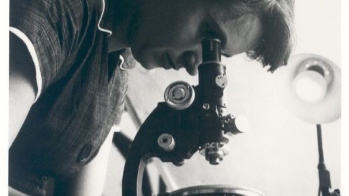
By the dawn of 1953, the question in molecular biology was no longer whether DNA was the hereditary material, but how its physical geometry could execute the twin imperatives of genetics: to store an astronomical volume of biological information, and to replicate that information with near-absolute fidelity during every cell division. At the Cavendish Laboratory in Cambridge and King’s College London, four individuals were entangled in an intense, competitive drama: James Watson (an ambitious twenty-four-year-old American ornithologist turned molecular biologist), Francis Crick (a brilliant thirty-six-year-old British physicist transitioning to biology), Maurice Wilkins (a biophysicist at King’s working on DNA fibers), and Rosalind Franklin (an extraordinarily gifted physical chemist and master of X-ray crystallography).
The Clues: Chargaff’s Rules and Photo 51
The structural puzzle rested upon three essential pillars of physical and chemical data:
- Chargaff’s Equimolar Rules (1950): Austro-Hungarian biochemist Erwin Chargaff had analyzed DNA across diverse species (from yeast to humans), discovering that regardless of base composition, the amount of adenine always equaled thymine ($A = T$), and guanine always equaled cytosine ($G = C$), such that the purines equaled the pyrimidines ($A + G = T + C = 50%$). Chargaff himself did not understand the structural significance of this rule.
- Franklin’s Photo 51: Working at King’s College, Rosalind Franklin discovered that DNA fibers existed in two distinct hydration states: the dry, crystalline “A” form, and the hydrated, paracrystalline “B” form. In May 1952, using an ultra-fine micro-camera and regulating humidity with hydrogen gas bubbled through salt solutions, Franklin exposed a single hydrated fiber of B-DNA to X-rays for 62 hours. The resulting diffraction pattern—immortalized as Photo 51—was of breathtaking clarity. It displayed a bold, cross-shaped pattern of dark reflections ($X$-shape) diagnostic of a helix, with ten layer lines per repeat cycle, an axial spacing of 3.4 Ångströms ($0.34text{ nm}$) between successive stacked bases, and a complete helical pitch of 34 Ångströms ($3.4text{ nm}$).
- The Antiparallel Monoclinic Lattice: Franklin’s crystallographic measurements revealed that DNA belonged to the monoclinic space group $C2$, possessing face-centered two-fold symmetry. When Crick examined Franklin’s data in an Medical Research Council (MRC) report shown to him by Max Perutz, his mathematical training in crystallography allowed him to grasp instantly what others had missed: the two complementary helical backbones must run in antiparallel directions—one running $5′ rightarrow 3’$, and the other running $3′ rightarrow 5’$.
The Cardboard Breakthrough in the Austin Wing
Linus Pauling at Caltech had mistakenly published a three-stranded DNA model with the phosphate backbones packed into the dense core and the bases radiating outward—a physical impossibility because the negatively charged phosphate oxygens would violently repel one another at physiological pH. Watson and Crick realized the hydrophilic sugar-phosphate backbones had to reside on the exterior facing the aqueous cellular cytoplasm, with the hydrophobic nitrogenous bases tucked inside.
On the morning of February 28, 1953, in the Austin Wing of the Cavendish Laboratory, Watson sat cutting cardboard representations of the four nitrogenous bases. At the suggestion of visiting American physical chemist Jerry Donohue, Watson abandoned the erroneous “enol” tautomeric forms depicted in standard organic textbooks and used the correct keto forms of guanine and thymine.
As Watson slid the cardboard cutouts across his desk, a revelation of breathtaking symmetry occurred:
- An adenine-thymine ($A-T$) pair joined by two specific hydrogen bonds possessed the exact same overall dimensions and geometry as a guanine-cytosine ($G-C$) pair joined by three hydrogen bonds.
- A bulky two-ring purine ($A$ or $G$) was always paired with a slender single-ring pyrimidine ($T$ or $C$), maintaining an invariant cross-sectional width of 20 Ångströms ($2.0text{ nm}$) along the entire length of the double helix.
“It has not escaped our notice that the specific pairing we have postulated immediately suggests a possible copying mechanism for the genetic material.”
— James Watson & Francis Crick, Nature (April 25, 1953)
Watson and Crick famously celebrated at The Eagle pub in Cambridge, announcing they had found “the secret of life.” In a single 900-word paper published in Nature on April 25, 1953, accompanied by companion papers from Franklin and Gosling and Wilkins, Watson and Crick unveiled the Antiparallel B-DNA Double Helix. The genetic code was written as a linear sequence of base pairs; and because each strand was the complementary mirror image of the other, unwinding the two strands provided two identical templates for replication. Biology had crossed into the era of molecular certainty.