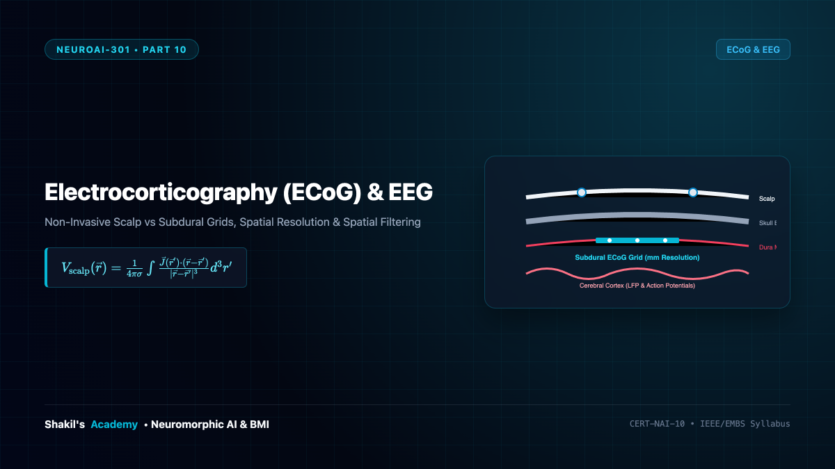
While penetrating intracortical microelectrodes provide the ultimate single-neuron resolution, their invasive nature carries surgical risks, chronic foreign-body tissue reactions, and limited spatial coverage. For many clinical brain-machine interfaces, recording aggregated field potentials from the cortical surface or the scalp offers an ideal compromise between safety, signal longevity, and information bandwidth. This part contrasts Electroencephalography (EEG) and Electrocorticography (ECoG), detailing spatial filtering algorithms and the pivotal role of High-Gamma Band (70–150 Hz) power.
1. Non-Invasive Scalp EEG and Volume Conduction
First recorded in humans by Hans Berger in 1924, Electroencephalography (EEG) records macroscopic electrical activity via non-invasive metal electrodes (e.g., ( ext{Ag/AgCl})) affixed to the scalp using conductive electrolyte gel:
- Signal Genesis: EEG does not measure single-unit action potentials (which are too brief and asynchronous to summate across distance). Instead, it measures the synchronized post-synaptic potentials (PSPs) of hundreds of thousands of spatially aligned pyramidal neurons oriented perpendicularly to the cortical surface.
- The Volume Conduction Smearing: Before reaching the scalp, electric currents must pass through the cerebral cortex ((sigma approx 0.33 ext{S/m})), cerebrospinal fluid (CSF, (sigma approx 1.79 ext{S/m})), skull bone ((sigma approx 0.006 ext{S/m})), and scalp skin ((sigma approx 0.43 ext{S/m})). The low-conductivity skull acts as a severe spatial low-pass filter:
[
ext{Spatial Resolution of EEG} approx 20 ext{ to } 30 ext{ mm}
]
High-frequency neural rhythms ((> 40 ext{ Hz})) are strongly attenuated, limiting typical scalp EEG analysis to classic low-frequency bands: Delta ((1 ext{–}4 ext{ Hz})), Theta ((4 ext{–}8 ext{ Hz})), Alpha ((8 ext{–}12 ext{ Hz})), and Beta ((13 ext{–}30 ext{ Hz})).
2. Subdural and Epidural Electrocorticography (ECoG)
Electrocorticography (ECoG)—also known as intracranial EEG (iEEG)—places platinum or gold disk electrode arrays directly onto the surface of the brain, either underneath the dura mater (subdural) or resting above the dura (epidural):
| Modality | Invasiveness | Spatial Resolution | Temporal Bandwidth | Typical Signal Amplitude |
|---|---|---|---|---|
| Scalp EEG | Completely Non-Invasive | (20 ext{ to } 30 ext{ mm}) | (0 ext{ to } 40 ext{ Hz}) | (10 ext{ to } 100 mu ext{V}) |
| Epidural ECoG | Craniotomy / Burr Hole | (3 ext{ to } 5 ext{ mm}) | (0 ext{ to } 200 ext{ Hz}) | (50 ext{ to } 200 mu ext{V}) |
| Subdural ECoG | Open Dura Craniotomy | (1 ext{ to } 2 ext{ mm}) | (0 ext{ to } 500 ext{ Hz}) | (100 ext{ to } 1000 mu ext{V}) |
| Intracortical Arrays | Parenchyma Penetration | (0.05 ext{ to } 0.1 ext{ mm}) | (0 ext{ to } 5000 ext{ Hz}) | (50 ext{ to } 500 mu ext{V}) |
By placing electrodes beneath the insulating skull bone, ECoG gains an order of magnitude higher spatial resolution and preserves ultra-fast cortical dynamics without penetrating the delicate cerebral parenchyma.
3. The Significance of High-Gamma Band Activity (70–150 Hz)
In human brain-machine interfaces, the High-Gamma Band (70 to 150 Hz) is the crown jewel of ECoG decoding:
- Unlike low-frequency alpha and beta rhythms (which reflect widespread, distributed inhibitory idling), local high-gamma power is strictly localized to active cortical columns ((< 2 ext{ mm}) radius).
- Empirical and biophysical studies (Crone, Miller, Chang) demonstrate that high-gamma envelope power (P_{ ext{HG}}(t)) correlates almost perfectly ((r > 0.85)) with the underlying multi-unit population firing rate:
[
P_{ ext{HG}}(t) propto sum_{k} ext{FR}_k(t)
] - High-gamma power provides the core feature for decoding speech acoustics, finger movements, and complex cognitive intentions with low latency.
4. Spatial Filtering: Surface Laplacian and Common Spatial Patterns
Because volume conduction produces cross-talk where multiple adjacent channels record identical deep dipole sources, mathematical spatial filtering is mandatory:
- Bipolar Derivation: Subtracts adjacent channel pairs: (V_i^{ ext{bipolar}} = V_i – V_{i+1}), canceling out common far-field ambient noise.
- Surface Laplacian (Current Source Density): Discretizes the 2D Laplace-Beltrami operator to compute local current entering or exiting the skull:
[abla^2 V_i approx rac{4}{d^2} left( V_i – rac{1}{4} sum_{j in ext{neighbors}} V_j
ight)
]
This dramatically sharpens the spatial focus of scalp EEG, isolating cortical generators. - Common Spatial Pattern (CSP): An optimal supervised spatial projection matrix (W) that maximizes the variance for one cognitive condition (e.g., right hand motor imagery) while simultaneously minimizing variance for a competing condition (e.g., left hand imagery):
[
J(w) = rac{w^T Sigma_1 w}{w^T Sigma_2 w}
]
solved via generalized eigenvalue decomposition (Sigma_1 w = lambda Sigma_2 w).