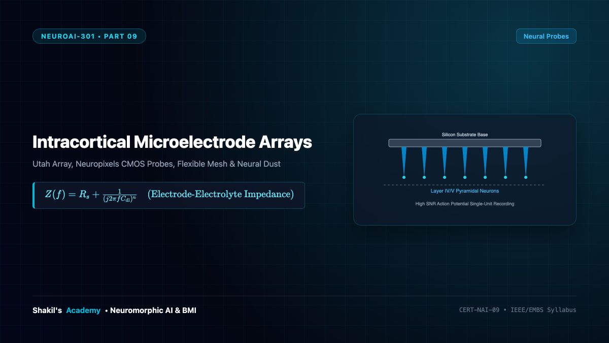
To decode intentions or thoughts directly from the central nervous system, brain-machine interfaces must interface with single neurons at single-millisecond and micrometer resolutions. Extracellular electrophysiology measures the tiny microvolt-scale ionic field fluctuations produced when an action potential propagates down an axon. This part explores the technological evolution of Intracortical Microelectrode Arrays: from the clinical gold-standard silicon Utah Array to high-density CMOS Neuropixels probes, biocompatible flexible polymer mesh threads, and wireless ultrasonic Neural Dust motes.
1. The Utah Array: Silicon Micromachining Pioneer
Invented by Richard Normann at the University of Utah in the late 1980s (commercialized by Blackrock Neurotech), the Utah Array remains the only intracortical array with extensive FDA Investigational Device Exemption (IDE) human clinical trial history (e.g., BrainGate consortium):
- Geometry: A (4 imes 4 ext{ mm}) monocrystalline silicon substrate supporting a (10 imes 10) grid of 100 micromachined needle-like electrodes (96 active recording channels).
- Dimensions: Electrodes are (1.0 ext{ to } 1.5 ext{ mm}) long with (400 mu ext{m}) pitch, engineered to penetrate through cortical Layer I to terminate directly in Layer IV/V, home to the large pyramidal motor output neurons.
- Material Science: The silicon shanks are insulated with polyimide or parylene-C, terminating in conductive platinum or iridium oxide (( ext{IrO}_x)) tips.
- Electrode Impedance: Tips are engineered for low electrochemical impedance ((Z approx 200 ext{ to } 800 ext{k}Omega) at (1 ext{ kHz})), maximizing the signal-to-noise ratio (SNR) for recording single-unit action potentials ((50 ext{ to } 500 mu ext{V})).
2. Neuropixels: The CMOS Revolution in High-Density Electrophysiology
Developed by HHMI Janelia, IMEC, and the Allen Institute (Jun et al., 2017), Neuropixels revolutionized neuroscience by integrating standard silicon CMOS fabrication directly onto neural recording shanks:
| Specification | Classic Utah Array | Neuropixels 1.0 (2017) | Neuropixels 2.0 (2021) |
|---|---|---|---|
| Recording Sites | 96 active channels | 960 titanium nitride sites | 5,120 sites across 4 shanks |
| Simultaneous Channels | 96 channels | 384 switchable channels | 768 switchable channels |
| Electrode Pitch | (400 mu ext{m}) | (20 mu ext{m}) (High density) | (15 mu ext{m}) staggered pitch |
| Shank Length & Width | (1.5 ext{ mm} imes 4 ext{ mm}) block | (10 ext{ mm} imes 70 mu ext{m}) | (10 ext{ mm} imes 70 mu ext{m}) |
| On-Chip Electronics | Passive wire bundle out | On-shank amplifiers, filters, ADCs | Full on-chip digital serialization |
A single 10 mm Neuropixels shank can span multiple brain regions simultaneously (e.g., from cerebral cortex, through hippocampus, down into the thalamus), recording thousands of single-unit spikes in freely moving subjects with zero external analog cabling.
3. Flexible Polymer Threads and Robotic Insertion (Neuralink)
A primary limitation of rigid silicon arrays is the dramatic mechanical mismatch between brittle silicon (Young’s modulus (E sim 150 ext{ GPa})) and soft biological brain parenchyma ((E sim 1 ext{ to } 3 ext{ kPa})). Micromotion caused by vascular pulsation and respiration induces constant shear stress, triggering chronic neuroinflammatory cascades and glial scarring that encapsulate electrodes within months.
To solve this, modern systems (such as Neuralink’s N1 device and Charles Lieber’s syringe-injectable mesh) employ ultra-flexible thin-film polymer substrates (polyimide or parylene, (E sim 2 ext{ to } 3 ext{ GPa})):
- Threads are just (4 ext{ to } 6 mu ext{m}) thick, flexing naturally with biological tissue and drastically suppressing astrocytic and microglial immune activation.
- Because flexible threads cannot penetrate tissue on their own, rapid automated stereotaxic surgical robots use ultra-fine tungsten insertion needles (vision-guided to avoid surface blood vessels) to insert threads at rates of dozens per minute.
4. Neural Dust: Wireless Ultrasonic Nanomotes
Developed by Maharbiz, Carmena, and Alon at UC Berkeley (2013–2016), Neural Dust eliminates transcutaneous wires and batteries altogether. Sub-millimeter implantable motes ((sim 0.8 ext{ mm}^3)) are deployed inside deep tissue:
- An external ultrasonic transceiver emits megahertz acoustic pulses ((f sim 1 ext{ to } 3 ext{ MHz})). Ultrasound penetrates biological tissue with orders-of-magnitude lower attenuation than electromagnetic radio waves ((0.5 ext{ to } 1 ext{ dB/cm/MHz}) vs severe RF absorption).
- A piezoelectric lead zirconate titanate (PZT) crystal on the dust mote harvests acoustic energy, converting it into electrical power.
- Extracellular action potentials modulate the gate of an onboard custom transistor, altering the electrical load across the piezoelectric crystal.
- This load change dynamically alters the acoustic reflection coefficient, scattering back an amplitude-modulated ultrasonic echo that the external transceiver decodes into neural voltage waveforms!