Electrical microelectrodes suffer from an inescapable physical limitation: electrical current spreads indiscriminately through tissue, exciting or inhibiting all cell types within a radius regardless of their physiological role (e.g., activating excitatory pyramidal neurons and inhibitory GABAergic interneurons simultaneously). In 2005, Karl Deisseroth, Ed Boyden, and Feng Zhang pioneered Optogenetics: genetically inserting light-sensitive microbial opsin ion channels into specific neural subpopulations. Combined with genetically encoded fluorescent indicators and focused ultrasound, optical and genetic interfaces provide cell-type-specific, single-neuron interrogation of brain circuits.
1. Microbial Rhodopsins: Channelrhodopsin-2 and Halorhodopsin
Optogenetics harnesses microbial opsin genes transferred into mammalian neurons via Adeno-Associated Viral (AAV) vectors under cell-type-specific promoters (e.g., CamKII(alpha) for excitatory neurons, PV or SOM for inhibitory interneurons):
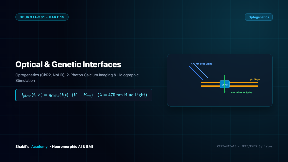
- Channelrhodopsin-2 (ChR2): Derived from the green alga Chlamydomonas reinhardtii, ChR2 is a light-gated cation channel. Upon illumination with blue light ((lambda approx 470 ext{ nm})), the all-trans-retinal chromophore photo-isomerizes to 13-cis-retinal, opening the pore to allow passive inward flux of Sodium (( ext{Na}^+)) and Calcium (( ext{Ca}^{2+})) ions:
[
I_{ ext{ChR2}}(t, V) = g_{ ext{ChR2}} cdot O(t) cdot (V – E_{ ext{rev}})
]
This depolarizes the membrane past threshold within milliseconds, driving precise, millisecond-locked action potential firing. - Halorhodopsin (NpHR): Cloned from the archaeon Natronomonas pharaonis, NpHR is an electrogenic chloride pump activated by yellow/amber light ((lambda approx 590 ext{ nm})). It actively pumps Chloride (( ext{Cl}^-)) ions into the cell, hyperpolarizing the neuron and silencing spike activity on demand.
By pairing blue ChR2 (optical ignition) and yellow NpHR (optical braking), researchers can turn targeted neural circuits on and off with single-spike precision.
2. Genetically Encoded Calcium Indicators: The GCaMP Family
While opsins write information into neurons, Genetically Encoded Calcium Indicators (GECIs) read information out optically. When an action potential fires, voltage-gated calcium channels open, causing a transient ten-fold surge in intracellular calcium concentration (from (approx 50 ext{ nM}) to (> 1 mu ext{M})).
The engineered biosensor GCaMP is a circular permutation of Green Fluorescent Protein (cpGFP) fused to Calmodulin (CaM) and the M13 peptide:
[
ext{Ca}^{2+} + ext{CaM-M13}
ightleftharpoons ext{Complex} implies ext{Fluorescence Jump } (Delta F / F_0 > 500%)
]
Binding of ( ext{Ca}^{2+}) induces a conformational shift that shields the fluorophore from water solvent quenching, causing a bright green fluorescent flash lasting (sim 100 ext{ ms}).
3. Two-Photon Laser Scanning Microscopy
Because biological brain tissue strongly scatters visible light ((l_s approx 50 mu ext{m})), single-photon widefield microscopy cannot image beneath the superficial cortex. Two-Photon Microscopy (Denk, Strickler, Webb, 1990) circumvents Rayleigh scattering by using pulsed near-infrared femtosecond lasers ((lambda approx 920 ext{ nm})):
- Infrared light experiences significantly reduced tissue scattering and absorption.
- Fluorescence excitation occurs only at the focal spot where the photon density is high enough for a fluorophore molecule to absorb two infrared photons simultaneously ((E = 2 h
u_{ ext{IR}} = h
u_{ ext{vis}})). - Because two-photon excitation scales quadratically with intensity ((I^2)), there is zero out-of-focus background fluorescence, allowing high-resolution imaging of thousands of individual dendritic spines up to (1 ext{ mm}) deep in intact, behaving brains.
4. Transcranial Focused Ultrasound Neuromodulation (tFUS)
For clinical applications where genetic viral vector delivery is ethically or regulatory restricted, Transcranial Focused Ultrasound (tFUS) provides a non-genetic, non-invasive alternative:
[
I_{ ext{SPPA}} sim 1 ext{ to } 50 ext{ W/cm}^2, quad f approx 250 ext{ to } 650 ext{ kHz}
]
A phased array transducer focuses acoustic waves through the intact human skull into deep subcortical nuclei (such as the thalamus or amygdala) with millimeter precision. Acoustic radiation forces and cavitation activate mechanosensitive ion channels (PIEZO1, TRP), exciting or suppressing deep brain circuits without requiring surgical craniotomy.