Thirty-four million years ago, at the Eocene-Oligocene transition, global temperatures plummeted, transforming the lush polar rainforests of the Eocene into ice sheets. In this cooling, seasonal world, the surviving anthropoid primates in Africa and Arabia underwent a decisive evolutionary bifurcation: the divergence of the Platyrrhini (flat-nosed New World monkeys, who rafted across a narrower Atlantic Ocean to South America) from our direct ancestral lineage, the Catarrhini (downward-facing nosed Old World monkeys and apes). Over the next twenty million years, Catarrhines achieved profound biological milestones: a genomic duplication that restored full trichromatic color vision, an ancient transposable element insertion that deleted the external tail, and an explosive Miocene adaptive radiation that forged the orthograde, suspensory body plan shared by all apes and humans.
1. The Opsin Gene Duplication: Reclaiming the Trichromatic World
As explored in Part 13, stem mammals sacrificed two color opsin genes ($SWS2$ and $Rh2$) during their 150-million-year Mesozoic nocturnal exile, reducing ancestral placental mammals to dichromatic vision (red-green colorblindness). For tens of millions of years, early primates navigated the canopy using only short-wavelength ($SWS1$, blue, ~420 nm) and long-wavelength ($LWS$, green-yellow, ~540 nm) photoreceptors.
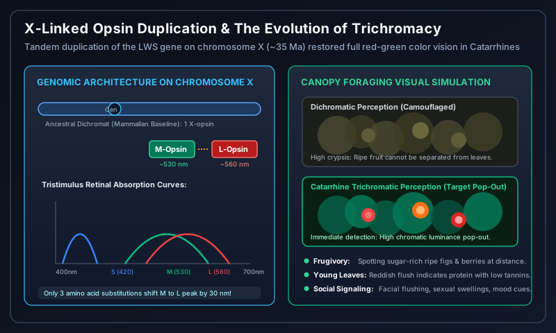
Approximately $35\text{–}30\text{ Ma}$, in the ancestral catarrhine lineage inhabiting the Fayum Depression of Egypt, an unequal homologous crossing-over event altered primate vision forever. The $LWS$ opsin locus on the X chromosome underwent a tandem gene duplication, creating two adjacent, nearly identical opsin genes:
The Five-Sites Rule and Spectral Tuning
Following duplication, the two tandem genes accumulated subtle point mutations. Groundbreaking genetic work by Jeremy Nathans and Gerald Jacobs demonstrated that just three to five critical amino acid substitutions (at positions 180, 277, 285, 308, and 309 inside the retinal-binding transmembrane pocket) shifted the peak absorption wavelength ($\lambda_{max}$) of one duplicated opsin by $30\text{ nm}$:
- The ancestral-derived M-Opsin (Medium-Wavelength Sensitive) retained peak sensitivity at $\lambda_{max} \approx 530\text{ nm}$ (Green).
- The newly diverged L-Opsin (Long-Wavelength Sensitive) shifted peak sensitivity to $\lambda_{max} \approx 560\text{ nm}$ (Red).
Working in conjunction with the autosomal short-wavelength opsin (S-Opsin, on Chromosome 7, $\lambda_{max} \approx 420\text{ nm}$), Catarrhine primates regained routine trichromacy. Unlike New World Platyrrhines (where trichromacy is polymorphic and mostly restricted to heterozygous females), every single male and female Old World monkey, ape, and human inherited two distinct X-linked opsin genes.
The Triple Selective Engine
- Frugivory (The Ripe Fruit Hypothesis): Ripe figs and fruits reflect wavelengths $>580\text{ nm}$. To a dichromat, red fruits blend seamlessly into green foliage. To a trichromat, red fruit triggers instant chromatic luminance “pop-out” across hundred-meter distances.
- Folivory (The Young Leaf Hypothesis): In seasonal tropical forests, mature leaves are saturated with indigestible cellulose and toxic tannins. Young, nutrient-rich leaves flush crimson and orange with protective anthocyanins. Trichromacy allowed Catarrhines to pinpoint high-protein edible leaves at a glance.
- Socio-Sexual Signaling: Trichromacy facilitated naked facial skin and sexual swellings that flush with oxygenated hemoglobin (e.g., baboon chests, human blushing), transforming color into a high-fidelity social communication channel.
2. The Genetic Mystery of Tail Loss: The TBXT Alu Insertion
For roughly 40 million years of primate history, every known fossil primate possessed a functional external tail. In tree-dwelling monkeys, the tail is an active biomechanical rudder, providing dynamic angular momentum balance during high-speed arboreal running and leaping. Yet by $25\text{ million years ago}$, in the common ancestor of all apes and humans (the superfamily Hominoidea), the external tail vanished entirely, reduced to an internal vestigial fused structure: the coccyx.
For decades, the genetic catalyst of this dramatic macroevolutionary transition remained a profound riddle. In February 2024, a landmark study published in Nature by Bo Xia, Jef Boeke, Itai Yanai, and their team uncovered the exact genomic smoking gun:
The Jumping Gene Mechanism
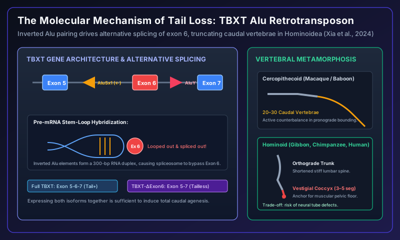
The TBXT gene (formerly known as Brachyury or T) encodes an ancient T-box transcription factor essential for embryonic notochord and caudal vertebral specification. The researchers discovered that approximately 25 Ma, an Alu mobile genetic element (AluY) hopped into intron 6 of the TBXT gene in a stem hominoid.
Crucially, an older primate Alu element (AluSx1) was already present in intron 5—oriented in the reverse (inverted) direction. When the pre-mRNA was transcribed:
- The two complementary Alu RNA sequences paired up, forming a tight, 300-base-pair double-stranded RNA stem-loop.
- This hairpin loop physically pinched out Exon 6, concealing its splice donor and acceptor sites from the cellular spliceosome.
- As a consequence, hominoid cells began producing two distinct splicing isoforms: the full-length transcript and a truncated isoform lacking exon 6 ($TBXT^{\Delta exon6}$).
When the researchers inserted this identical Alu element into genetically engineered mice, the mice were born completely tailless! The simultaneous expression of both isoforms disrupts caudal somite elongation just enough to terminate tail growth while leaving normal spinal cord development intact.
The Biomechanical Trade-Off
Why did natural selection preserve tail loss? In an upright, climbing ape, a dangling tail offered minimal counterbalance while increasing body mass and drag. Taillessness enabled the evolution of a reinforced, muscular pelvic floor (the levator ani complex), capable of supporting visceral organ weight during orthograde (vertical) posture. However, this evolutionary leap came at a cost: the authors noted that mice expressing $TBXT^{\Delta exon6}$ exhibited increased susceptibility to neural tube defects (spina bifida)—an evolutionary scar preserved in human medical epidemiology to this day.
3. The Miocene Ape Paradise: Orthogrady and the Y-5 Molar Pattern
During the Miocene epoch ($23\text{–}5.3\text{ Ma}$), the world experienced the true Golden Age of Apes. While today only five genera of hominoids survive (gibbons, orangutans, gorillas, chimpanzees, and humans), the Miocene Old World was teeming with ape diversity: over 40 extinct genera and 100 species flourished across Africa, Europe, and Asia.
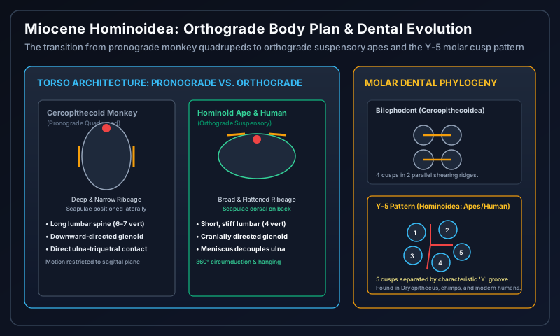
The Orthograde Biomechanical Revolution
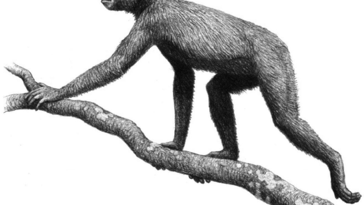
Basal Miocene apes like Proconsul ($20\text{ Ma}$) in Kenya still walked along the tops of branches on all fours (pronograde arboreal quadrupedalism). But as the early Miocene progressed into the middle and late Miocene, taxa such as Morotopithecus, Pierolapithecus, Dryopithecus, and Hispanopithecus dismantled the monkey quadrupedal body plan, constructing the orthograde (upright) suspensory Bauplan:
- Broad, Shallow Ribcage: Monkeys have a laterally compressed, deep ribcage that keeps limbs directly beneath the body for galloping. Apes widened the thorax dorsoventrally, moving the center of gravity backward toward the vertebral column.
- Dorsal Scapulae and Cranial Glenoid Fossa: The shoulder blades migrated from the sides of the chest onto the back (dorsal surface). The shoulder socket (glenoid fossa) angled upward, granting the arm a full $360^\circ$ range of motion and enabling below-branch arm hanging (brachiation and clambering).
- Decoupled Ulnar Wrist Meniscus: In monkeys, the ulna articulates directly with the triquetral bone of the wrist, locking the hand in a stable walking posture. In hominoids, the ulna retreated, replaced by a fibrocartilaginous meniscus disc. This granted the ape wrist extraordinary degrees of adduction, abduction, and rotation.
- Shortened Lumbar Spine: Monkeys have 6 to 7 flexible lumbar vertebrae for bounding. Apes reduced their lumbar vertebrae to 4 or 5, stiffening the lower back to resist mechanical buckling during upright climbing.
The Dental Signature: The Y-5 Molar Pattern
In the fossil record, where postcranial skeletons are often lost to time, paleontologists diagnose hominoids through dental morphology. While Old World Cercopithecoid monkeys evolved bilophodont molars (four cusps connected in two parallel shearing ridges, specialized for masticating tough leaves), all hominoids retain the ancestral Y-5 Molar Pattern:
- The lower molar possesses five distinct cusps (protoconid, metaconid, hypoconid, entoconid, and hypoconulid).
- The valleys between cusps 2 and 3 form an unmistakable “Y”-shaped groove system.
From 12-million-year-old Dryopithecus in Europe to Sivapithecus in Pakistan, to modern chimpanzees and the teeth in your own lower jaw, the Y-5 molar remains an indelible morphological signature of our Miocene ape ancestry.
4. The Late Miocene Bottleneck: The African Stage is Set
Between 10 and 6 million years ago, the Miocene paradise ended. Continental drift closed the Tethys Seaway, the Tibetan Plateau surged upward, and the East African Rift system began to tear eastern Africa apart. Global climates grew colder and drier.
The vast subtropical forests of Europe and Asia withered into open temperate woodlands. The magnificent Eurasian apes—Dryopithecus, Ouranopithecus, Lufengpithecus, and the giant 3-meter-tall Gigantopithecus—faced catastrophic extinctions. In Africa, the unbroken equatorial rainforest fractured into a mosaic of gallery forests, open woodlands, and savanna grasslands.
Trapped in this changing African mosaic, a small, resilient population of orthograde, tailless, large-brained, trichromatic apes stood at the threshold of a new ecological frontier. As the trees grew further apart, walking between them became a matter of life and death. The stage was set for the Great Ape Split and the emergence of habitual bipedalism.