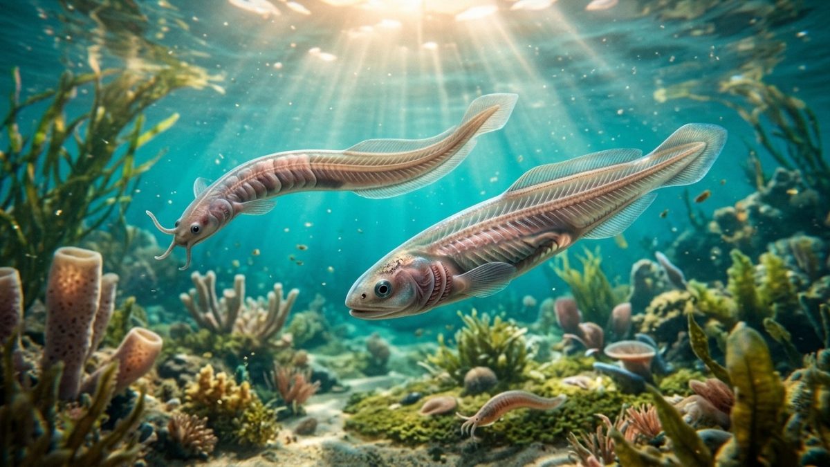
More than 525 million years ago, in the shadowed shallows of the Cambrian oceans, an unassuming ribbon of flesh scarcely longer than a human thumbnail embarked on one of the most consequential anatomical journeys in the history of life. Amidst a seabed dominated by armored arthropods and predatory priapulid worms, this creature possessed neither a mineral shell nor crushing jaws. Yet inside its body ran a flexible, fluid-filled rod of pressurized cells that would become the mechanical blueprint for every fish, amphibian, reptile, bird, and mammal that ever lived: the notochord. This is the story of Phylum Chordata and the developmental revolution that forged the vertebrate brain, face, and skeleton.
1. The Five Definitive Chordate Synapomorphies
To understand the evolutionary genesis of humanity, we must trace our lineage back to the deuterostome branch of bilaterian animals. While protostomes (such as arthropods, annelids, and mollusks) develop their mouth from the primary embryonic blastopore and position their main nerve cords ventrally along the belly, deuterostomes (chordates, hemichordates, and echinoderms) develop the blastopore into the anus, forming the mouth secondarily, and run their central nervous system along the dorsal spine.
Every member of our phylum—from the translucent marine lancelet (Branchiostoma) to modern Homo sapiens—is defined by five homologous anatomical innovations present at some stage of embryonic development:
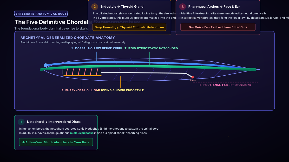
- The Notochord: A flexible, hydrostatic axial rod composed of vacuolated, glycoprotein-filled cells wrapped inside tightly woven collagenous sheaths. It prevents the body from shortening during lateral muscle contractions, translating lateral wave-like undulations into forward thrust. In adult humans, embryonic notochord remnants persist as the cushion-like nucleus pulposus within our intervertebral discs.
- The Dorsal Hollow Nerve Cord: Formed through primary neurulation by the invagination and folding of dorsal ectoderm. In non-chordate invertebrates, nerve cords are solid and ventral; in chordates, the dorsal cord envelops a fluid-filled neurocoel, which later enlarges anteriorly into the tripartite brain (prosencephalon, mesencephalon, and rhombencephalon).
- Pharyngeal Slits / Clefts: A series of bilateral lateral openings perforating the pharynx wall. Initially functioning as passive water-straining sieves for filter feeding, they transformed into gas-exchange gills in aquatic vertebrates and into Eustachian tubes, middle ear cavities, and parathyroid glands in land-dwelling tetrapods.
- The Endostyle (Thyroid Precursor): A ciliated, glandular groove along the ventral floor of the pharynx that secretes mucus to trap microscopic food particles. Critically, its cells concentrate environmental iodine to synthesize mono- and diiodotyrosine—marking it as the direct homologous evolutionary precursor to the vertebrate thyroid gland.
- The Post-Anal Tail: An active muscular elongation extending posterior to the digestive terminus (anus). Flanked by segmented, V-shaped or W-shaped myomeres, it transformed the posterior body into a dedicated locomotive hydrofoil.
2. Neural Crest Cells: The Developmental Miracle of the ‘New Head’
While basal chordates like amphioxus possess the five foundational structures, they remain blind, brainless, and passive filter-feeders that lack a true skull, camera-style sensory eyes, or predatory teeth. What miraculous developmental leap converted sluggish mud-burrowers into active, hunting vertebrates?
In 1983, evolutionary morphologists Carl Gans and R. Glenn Northcutt formulated the groundbreaking “New Head” Hypothesis. They revealed that the vertebrate revolution was driven by the emergence of a transient, pluripotential population of migratory embryonic cells so unique they are universally recognized today as the fourth germ layer: the Neural Crest Cells (NCCs).
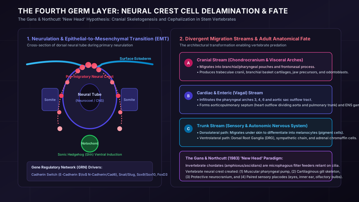
During early embryogenesis, as the neural plate folds to create the dorsal neural tube, cells at the neural plate border undergo an Epithelial-to-Mesenchymal Transition (EMT). Governed by a conserved gene regulatory network—including transcription factors Snail, Slug, Sox9, Sox10, and FoxD3—these cells downregulate tight adhesion junctions ($E\text{-cadherin}$ gives way to $N\text{-cadherin}$ and cadherin-6), detach from the neuroepithelium, and embark on extensive migratory voyages throughout the embryo:
- Cranial Neural Crest: Streams ventrally into the developing frontonasal prominence and pharyngeal arches. Unlike typical embryonic mesoderm, these cranial crest cells possess the remarkable capacity to form mineralized skeletal tissues—giving rise to the chondrocranium (cranial vault), trabeculae cranii, visceral jaw cartilages, sensory capsules around the eyes and ears, and dental odontoblasts that deposit dentine.
- Cardiac & Vagal Neural Crest: Colonizes the pharyngeal pouches and the aortic outflow tract of the heart, orchestrating the formation of the spiral aorticopulmonary septum that divides arterial blood from venous return. It also seeds the entire enteric nervous system (“the second brain” within the gut wall).
- Trunk Neural Crest: Migrates along two distinct routes:
- Dorsolateral pathway: Infiltrates the epidermis to differentiate into melanocytes, granting vertebrates protective camouflage, sexual signaling, and UV shields.
- Ventrolateral pathway: Penetrates through the anterior sclerotome to form the sensory Dorsal Root Ganglia (DRG), the sympathetic nervous chains, and the epinephrine-producing chromaffin cells of the adrenal medulla.
Without neural crest cells, vertebrates would have no skull to shield their expanding forebrain, no sensory capsules to focus stereoscopic vision and sound, no autonomic nervous system to orchestrate the flight-or-fight response, and no cartilaginous pharyngeal pump to replace sluggish cilia with high-volume, muscle-driven respiration.
3. The Fossil Record: Pikaia, Haikouichthys, and the Agnathans
The transition from invertebrate chordate to crown vertebrate is spectacularly documented across two of the world’s most pristine Cambrian Lagerstätten: the 508-million-year-old Burgess Shale in British Columbia, Canada, and the 518-million-year-old Chengjiang Biota in Yunnan Province, China.
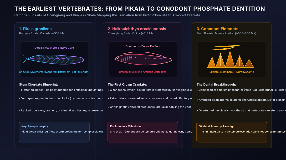
Pikaia gracilens: The Proto-Chordate Prototype
Discovered by Charles Walcott in 1911 and re-analyzed in groundbreaking detail by Simon Conway Morris and Jean-Bernard Caron in 2012, Pikaia gracilens was a 4-centimetre-long, laterally flattened swimmer. It possessed a dorsal notochord extending almost to the anterior tip, approximately 100 distinct zigzagging muscle blocks (myomeres), and a dorsal blood vessel. However, Pikaia lacked a distinct head, eyes, and mineralized tissues, representing a stem chordate grade closely allied with living cephalochordates.
Haikouichthys ercaicunensis: The First True Craniate
Unearthed in the slightly older Chengjiang shales of Yunnan by Chinese paleontologist Shu Degan and colleagues in 1999, Haikouichthys ercaicunensis pushed the origin of true vertebrates back 518 million years—deep into the early Cambrian. Unlike Pikaia, Haikouichthys is an unambiguous craniate:
- It possessed a distinct cartilaginous head capsule housing a lobed anterior brain.
- It featured prominent paired lateral eyes and olfactory or otic sensory capsules.
- It displayed six to nine pairs of gill pouches supported by a branchial basket.
- Crucially, flanking its flexible notochord were microscopic repeated cartilaginous elements called arcualia—the evolutionary precursors to neural arches and vertebral segments.
Haikouichthys and its contemporary relative Myllokunmingia demonstrate that active, cephalized vertebrates armed with sensory optics and proto-vertebrae had already evolved alongside monstrous predators like Anomalocaris, playing an active locomotive role in the Cambrian explosion.
4. The Conodont Secret: Teeth Before Armor
For more than a century, an enduring dogma of vertebrate paleontology held that our internal mineralized skeleton evolved first as protective dermal armor—thick, bony plates encasing the exterior of jawless fish (ostracoderms) to shield them from giant predatory sea scorpions (eurypterids).
This “Armor First” hypothesis was decisively demolished by microscopic, amber-colored fossils known as conodonts. Ranging in size from 0.1 mm to several millimeters, conodont elements were discovered in vast quantities in marine strata from the Late Cambrian through the Triassic ($500\text{–}200\text{ Ma}$), but their biological identity remained an absolute enigma for over 130 years.
In 1983, a complete soft-bodied conodont animal was finally discovered in Carboniferous rocks near Edinburgh, Scotland. It was a slender, eel-like chordate equipped with a notochord, chevron myomeres, and giant camera eyes. At the front of its throat lay the famous conodont elements: an intricate, multi-component feeding apparatus consisting of delicate, needle-like grasping elements ($S$ and $M$ elements) and crushing, comb-like shearing plates ($P$ elements).
High-resolution crystallographic and histological analysis revealed the stunning reality: conodont elements are composed of genuine biomineralized calcium phosphate / hydroxyapatite: $$\text{Ca}_5(\text{PO}_4)_3(\text{OH})$$ Their microstructure displays enamel-like crowns and cellular dentine-like bases, showing clear microscopic wear facets from grinding, grasping, and slicing soft-bodied prey.
The evolutionary implication is profound: vertebrates invented biomineralized skeletal tissues not to defend themselves against predators, but to bite, slice, and hunt. Dental tissue preceded skeletal bone armor. Armed with a flexible notochord, a neural-crest-derived sensory skull, and phosphate slicing tools, the stage was set for our aquatic ancestors to take over the world’s oceans—setting the stage for the invention of the true, articulated jaw.