For the first two billion years of planetary history, life on Earth remained stubbornly microscopic. Bacteria and archaea radiated into every conceivable thermodynamic niche, mastering photosynthesis, nitrogen fixation, and methanogenesis, yet structurally they never transcended the basic prokaryotic body plan: tiny, wall-bound sacks of biochemistry. Then, around 1.8 billion years ago, an unprecedented endosymbiotic union occurred between two utterly divergent microbes—an event so biologically singular that it happened only once in four billion years, dissolving the bioenergetic ceiling of the prokaryotic cell and giving birth to every amoeba, redwood, mushroom, and human being on Earth.
1. The Asgard Archaea: Discovering Our True Ancestral Roots
For decades following Carl Woese’s classic ribosomal RNA phylogenies in 1977, biology taught the “Three-Domain Tree of Life”: Bacteria, Archaea, and Eukaryota diverging as three co-equal primary lineages from LUCA. However, this model left the morphological origin of eukaryotes an unresolved mystery—complex cells possess an internal cytoskeleton, endomembrane networks, and vesicular trafficking systems completely absent in typical bacteria and archaea.
In 2015, a revolutionary metagenomic discovery off the coast of Svalbard, Norway, shattered the three-domain dogma. A team led by Thijs Ettema recovered environmental DNA from deep hydrothermal sediment at Loki’s Castle and assembled the genome of an uncultivated organism named Lokiarchaeum. Subsequent expeditions identified an entire deep-branching superphylum: the Asgard Archaea (including Thor, Odin, and Heimdallarchaeota).
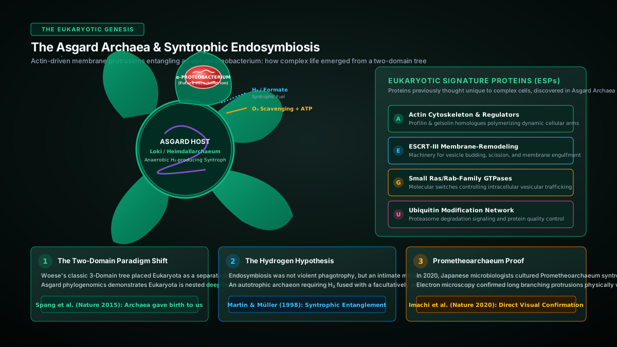
The Asgard genomes stunned the biological community. Embedded inside these archaea were dozens of genes previously believed to be unique to eukaryotes: Eukaryotic Signature Proteins (ESPs). They encoded authentic actin cytoskeletons, actin-binding regulators (profilin and gelsolin), small Ras-family GTPases for intracellular trafficking, and the entire ESCRT-III protein machinery responsible for vesicle budding and membrane remodeling.
Phylogenomic trees constructed with hundreds of conserved proteins confirmed that Eukaryotes do not form a third independent domain: we are branch of Archaea, emerging directly from within the Asgard superphylum as sister to Heimdallarchaeota.
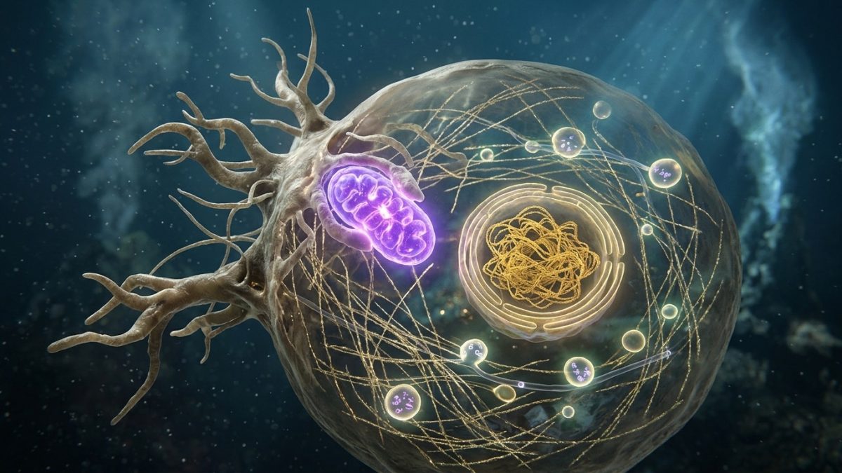
In 2020, Japanese microbiologist Masaru Nobu and colleagues achieved what was thought impossible: after 12 years of painstaking laboratory cultivation, they successfully isolated and photographed the first live Asgard archaeon, Prometheoarchaeum syntrophicum. Cryo-electron tomography revealed that rather than engulfing prey through phagocytosis, the cell produces long, delicate, branching tentacles that physically entangle partner bacteria, confirming the Hydrogen Hypothesis of Martin and Müller (1998): endosymbiosis was an intimate metabolic syntrophy where an anaerobic archaeon feeding on hydrogen embraced a facultatively aerobic, hydrogen-excreting $\alpha$-proteobacterium.
2. Mitochondriogenesis and the Bioenergetic Scaling Law
Why did complex life never evolve in bacteria? Why did bacteria remain small and morphologically simple despite experiencing four billion years of continuous natural selection and possessing immense biochemical diversity?
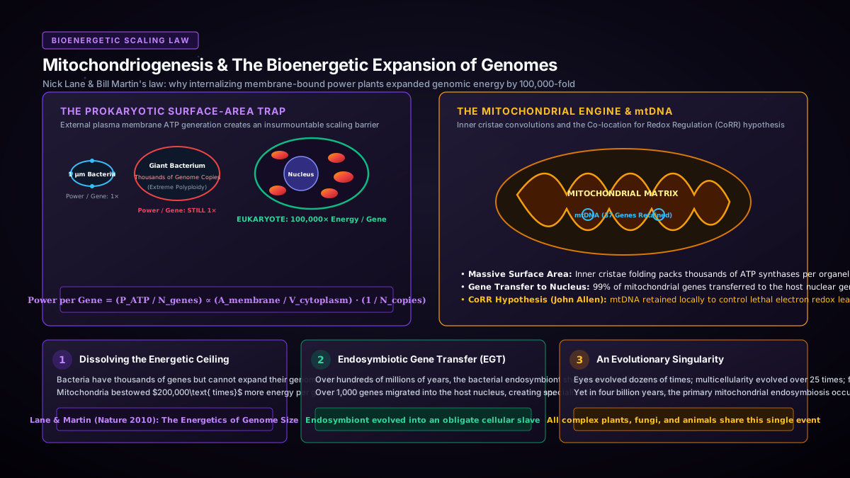
The answer, formulated by biochemist Nick Lane and evolutionary biologist William Martin in their groundbreaking 2010 Nature paper, is purely physical: bioenergetic scaling constraints.
Every bacterium generates its chemiosmotic proton gradient across its outer boundary: the plasma membrane. As a spherical or rod-shaped cell grows larger, its surface area expands with the square of its radius ($A \propto r^2$), while its internal cytoplasmic volume expands with the cube of its radius ($V \propto r^3$). Because synthesizing proteins to support a larger volume requires thousands of active ribosomes and continuous transcription, a large prokaryote faces an acute energy crisis:
$$\text{Power per Gene} = \frac{P_{\text{ATP}}}{N_{\text{genes}}} \propto \frac{A_{\text{membrane}}}{V_{\text{cytoplasm}}} \cdot \frac{1}{N_{\text{genome copies}}}$$When rare “giant bacteria” evolve—such as the $600\text{-}\mu\text{m}$ surgeonfish gut symbiont Epulopiscium fishelsoni or the sulfur pearl Thiomargarita magnifica—they cannot support this volume with a single enlarged genome. Instead, they must maintain up to tens of thousands of identical copies of their complete circular chromosome plastered against their outer membrane to control local respiration. Consequently, the energy available per individual gene copy in a giant bacterium is completely unchanged compared to a microscopic $1\text{-}\mu\text{m}$ E. coli.
The acquisition of the mitochondrion dissolved this energetic straitjacket forever. By internalizing the respiration-competent $\alpha$-proteobacterium, the host cell transformed its energy generation from a two-dimensional surface boundary into a three-dimensional internal network. Each mitochondrion folded its inner membrane into thousands of convoluted cristae, packing astronomical densities of electron transport complexes and ATP synthases inside the cell without compromising the host’s surface-to-volume ratio.
Furthermore, over hundreds of millions of years, the intracellular endosymbionts underwent massive genome reduction: non-essential genes were deleted or transferred to the host nucleus via Endosymbiotic Gene Transfer (EGT). Today, human mitochondria retain a tiny circular genome of only 37 genes. By delegating genetic administrative overhead to the central host nucleus while retaining miniature local genomes solely to control lethal electron redox leaks (the CoRR hypothesis of John Allen), the eukaryotic cell gained over $100,000\text{ to }200,000\text{ times}$ more ATP energy per gene than any bacterium on Earth. This energetic windfall provided the genomic budget required to evolve thousands of new regulatory genes, gene duplicates, complex signal transduction cascades, and macroscopic multicellular bodies.
3. The Genesis of the Nucleus and the Intron Defense Shield
While the mitochondrion solved the cell’s energetic crisis, its initial acquisition triggered an existential genetic catastrophe: the invasion of selfish mobile genetic elements.
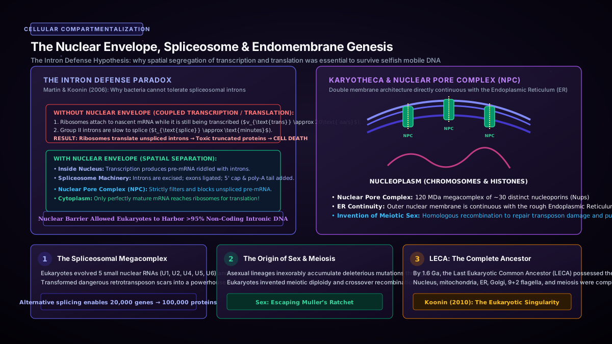
The ancestral $\alpha$-proteobacterium harbored thousands of Group II self-splicing introns—selfish retrotransposons capable of reverse-transcribing and copying themselves randomly throughout genomes. When endosymbionts periodically lysed inside the archaeal host, fragments of bacterial DNA integrated into the host’s chromosome, peppering vital archaeal genes with hundreds of parasitic mobile introns.
In prokaryotes, transcription and translation are obligatorily coupled: ribosomes physically attach to nascent mRNA while it is still being synthesized by RNA polymerase ($v_{\text{translation}} \approx 20\text{ amino acids/second}$). Splicing of an intron, however, is a slow catalytic process requiring minutes. In a coupled prokaryotic cytoplasm, ribosomes would inevitably read through unspliced introns before they could excise themselves, encountering in-frame stop codons and producing toxic truncated peptide fragments that would poison the cell.
To survive this genomic onslaught, the host cell evolved the single most defining morphological structure of our lineage: the nuclear envelope (karyotheca).
By folding endomembranes into a double lipid bilayer perforated by giant 120-MDa Nuclear Pore Complexes (NPCs), the cell created an absolute spatial and temporal barrier. Transcription and spliceosomal intron excision were confined exclusively to the interior of the nucleus. The nuclear pore complex acted as a strict molecular customs gate: only completely processed, spliced mRNA molecules bearing a $5’\text{-cap}$ and a $3’\text{-poly(A)}$ tail were permitted to exit into the cytoplasm for translation.
The evolutionary repurposing of this crisis was monumental: the host evolved the spliceosome—a massive RNA-protein machine composed of five small nuclear RNAs (U1, U2, U4, U5, U6) and over 150 proteins. Introns, once lethal genomic parasites, became the primary fuel for alternative splicing, allowing a single gene to encode dozens of distinct protein isoforms and conferring unparalleled proteomic flexibility on eukaryotic organisms.
Conclusion: The Emergence of LECA
By approximately $1.6\text{ billion years ago}$, all these transformative innovations had coalesced into a single ancestral population: the Last Eukaryotic Common Ancestor (LECA). LECA was not a primitive transition form; it was an extraordinarily sophisticated, fully modern eukaryotic cell equipped with:
- Mitochondria possessing functional cristae and ATP export mechanisms;
- A compartmentalized nucleus with nuclear pore complexes;
- A dynamic endomembrane system (Endoplasmic Reticulum, Golgi apparatus, lysosomes);
- A flexible actin-tubulin cytoskeleton with motor proteins (kinesin, dynein, myosin);
- A 9+2 microtubule axoneme forming the eukaryotic cilium / flagellum;
- And regular meiotic sex—diploid homologous chromosome pairing and crossover recombination to purge accumulated deleterious mutations and escape Muller’s ratchet.
From the crucible of LECA, the eukaryotic tree exploded into its major supergroups: Opisthokonta (animals and fungi), Archaeplastida (plants and red/green algae), SAR (stramenopiles, alveolates, rhizarians), and Amoebozoa. In Part 5, we will cross into Phase II and witness the momentous transition from solitary cells to cooperative multi-cellular organisms and the enigmatic macroscopic garden of the Ediacaran biota.
Key Academic Citations
- Spang, A., et al. (2015). Complex archaea that bridge the gap between prokaryotes and eukaryotes. Nature, 521(7551), 173–179.
- Imachi, H., et al. (2020). Isolation of an archaeon at the interface of the prokaryote–eukaryote divide. Nature, 577(7791), 519–525.
- Lane, N., & Martin, W. (2010). The energetics of genome complexity. Nature, 467(7318), 929–934.
- Martin, W., & Müller, M. (1998). The hydrogen hypothesis for the first eukaryote. Nature, 392(6671), 37–41.
- Martin, W., & Koonin, E. V. (2006). Introns and the origin of nucleus-cytosol compartmentalization. Nature, 440(7080), 41–45.
- Allen, J. F. (2003). Why chloroplasts and mitochondria contain genomes. Comparative and Functional Genomics, 4(1), 31–36.