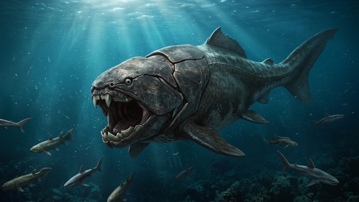
For more than one hundred million years after the first chordates undulated through Cambrian waters, vertebrates remained modest, bottom-dwelling mud-suckers confined to the lower tiers of the oceanic food pyramid. Giant sea scorpions (eurypterids) and shelled cephalopods ruled the Silurian seas. But between 440 and 380 million years ago, an astonishing morphological transformation re-engineered the front of the vertebrate gut into the most lethal anatomical weapon in biological history: the articulated jaw. With this single innovation, the Devonian Period exploded into the “Age of Fishes,” unleashing nine-metre armored apex predators like Dunkleosteus and bifurcating our aquatic ancestors into ray-finned swimmers and the lobe-finned pioneers destined to walk upon land.
1. The Respiratory Origins of Jaws: Homology of Branchial Arches
For decades, popular imagination assumed that jaws arose directly through selection for biting and predation. However, comparative embryology and functional morphology revealed an even more elegant evolutionary truth: our jaws did not evolve for eating—they evolved for breathing.
In basal jawless fishes (agnathans), the pharynx is supported by a series of repeating, cartilaginous visceral branchial arches (labeled 1 through 7) that keep the gill slits open. As these animals developed higher metabolic rates, relying on passive water flow across the gills became insufficient. In 1996, anatomist John Mallatt demonstrated that the anterior-most branchial arch (Arch 1) underwent massive muscularization to act as a high-powered ventilatory pump, forcefully expanding and contracting to flush oxygenated water across the downstream gills.
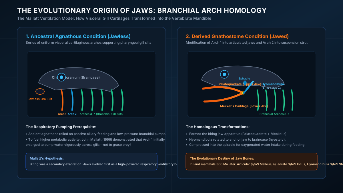
Once this powerful muscular hinge existed for respiratory pumping, natural selection swiftly co-opted it for grasping prey—a classic evolutionary exaptation. The precise morphological remodeling unfolded in three distinct steps:
- The Mandibular Arch (Arch 1): Folded forward and enlarged dramatically. Its dorsal half ossified into the palatoquadrate cartilage (the primary upper jaw, fusing to the neurocranium), while its ventral half formed Meckel’s cartilage (the structural core of the lower jaw/mandible).
- The Hyoid Arch (Arch 2): Rotated anteriorly to serve as an architectural anchor. Its dorsal element, the hyomandibula, functioned as an articulated brace wedged between the otic capsule of the braincase and the mandibular joint (a suspension mechanism known as hyostyly).
- Reduction to the Spiracle: The original first pharyngeal gill slit between Arch 1 and Arch 2 was compressed into a dorsal accessory respiratory opening called the spiracle (still visible today behind the eyes of rays and sharks), allowing bottom-dwelling bottom-feeders to inhale clean water while buried in sediment.
The Deep Homological Echo: In one of evolution’s most poetic transformations, when the synapsid ancestors of mammals remodeled their jaw joint 300 million years later, Meckel’s articular bone transformed into our middle ear malleus (hammer), the palatoquadrate’s quadrate bone became our incus (anvil), and the hyomandibula became our stapes (stirrup). Every time you hear music, you are listening through the repurposed gill arches of a Silurian fish!
2. Biomechanics of Dunkleosteus: The Four-Bar Linkage Monster
Armed with jaws, gnathostomes radiated into diverse lineages during the Devonian Period, none more terrifying than the Placoderms—armored fishes encased in massive bony thoracic shields. The undisputed apex titan of this era was Dunkleosteus terrelli, a monstrous predator reaching up to 9 metres in length and weighing over 4 metric tons.
Unlike modern sharks or bony fishes, Dunkleosteus possessed no true teeth rooted in enamel. Instead, its jaws were armed with self-sharpening, serrated dermal bone plates: the upper superognathal and lower inferognathal bones. Histological cross-sections reveal that these gnathal plates constantly sheared past each other during closure, utilizing abrasive friction to hone their cutting edges into razor-sharp scythes.
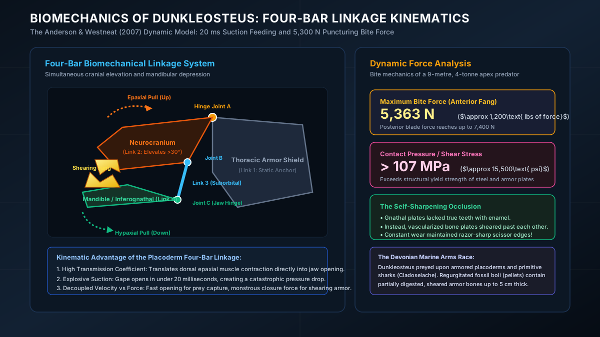
In 2007, biomechanical paleontologists Philip Anderson and Mark Westneat constructed a dynamic kinematic computer model of the Dunkleosteus skull, revealing that its jaw operated via a sophisticated planar four-bar mechanical linkage:
- Link 1 (Fixed Base): The massive dorsal thoracic armor shield.
- Link 2 (Driver): The neurocranium / head shield, hinged to the thoracic shield via bilateral ball-and-socket joints (Joint A). When massive dorsal epaxial muscles contracted, the entire head rotated upward by more than $30^\circ$.
- Link 3 (Transmission): The rigid suborbital plate connecting the skull to the lower jaw.
- Link 4 (Output): The inferognathal lower jaw, which depressed downward in synchronization with cranial elevation.
This four-bar linkage gave Dunkleosteus a dual mechanical superpower. In the opening phase, the system exhibited an extremely high velocity transmission coefficient: the monstrous mouth snapped open in less than 20 milliseconds ($0.02\text{ seconds}$), generating a catastrophic suction vacuum that pulled large fish and cephalopods directly into its maw.
In the closing phase, the linkage converted the mechanical leverage of immense jaw adductor muscles into staggering crushing power: $$F_{\text{bite}} \approx 5,363\text{ N} \quad (\approx 1,200\text{ lbs of force})$$ Because this monstrous force was concentrated upon the tiny contact area ($A \approx 0.5\text{ cm}^2$) of the anterior fangs, the puncturing pressure reached astronomical levels: $$P = \frac{F}{A} > 107\text{ MPa} \quad (\approx 15,500\text{ psi})$$ This contact pressure exceeded the structural yield strength of hardened steel and enabled Dunkleosteus to shatter through the thickest dermal bone plates of competing placoderms and primitive sharks like Cladoselache with effortless ease.
3. The Great Bifurcation: Ray-Fins (Actinopterygii) vs. Lobe-Fins (Sarcopterygii)
While placoderms dominated the open seas, another group of jawed vertebrates—the Osteichthyes (bony fishes)—underwent a fundamental phylogenetic divergence that would forever divide vertebrate destiny. Sometime in the Late Silurian, bony fishes split into two distinct subclasses based on the internal skeletal architecture of their fins:
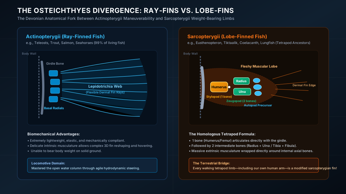
Subclass Actinopterygii: The Masters of Hydrodynamics
The ray-finned fishes confined their skeletal support bones (basal radials) entirely inside the body wall. The projecting fin itself is supported by an array of parallel, segmented, bony lepidotrichia (dermal fin rays) connected by delicate elastic webbing. This design is mechanically compliant, ultra-lightweight, and capable of undulating 3D curvature, granting ray-finned fishes unmatched maneuverability and acceleration. Today, Actinopterygii encompasses over 30,000 species—accounting for 99% of all living fish on Earth.
Subclass Sarcopterygii: The Architects of Walking
In contrast, the lobe-finned fishes adopted a radically different structural design. Instead of an internal fan of radials, their fin projected outward from the flank as a robust, fleshy muscular stalk supported by an internal, articulated chain of endochondral bones:
- The Monobasic Articulation: A single large proximal bone articulated directly with the pectoral girdle: the Humerus (or Femur in the pelvic fin).
- The Intermediate Zeugopod: The humerus articulated with two distal companion bones: the Radius and Ulna (or Tibia and Fibula).
- Extrinsic Musculature: Massive propulsive muscles wrapped directly around this axial bony core outside the main torso wall.
To an animal living in open pelagic water, a heavy, muscular lobe-fin was hydrodynamically inefficient compared to the feather-light ray-fin. But in the choked, weed-choked shallows of Devonian swamps and braided river deltas, the sarcopterygian lobe-fin was an extraordinary mechanical advantage. It allowed fish like Eusthenopteron and Panderichthys to prop themselves up on the riverbed, clamber over submerged logs, and crawl between evaporating pools.
4. The Lungs That Preceded Land: The Swim Bladder Story
A widespread misconception is that lungs evolved when vertebrates crawled onto land. In reality, molecular phylogenetics and paleontology prove the exact opposite: lungs evolved in water millions of years before any vertebrate set foot on land.
During the warm, sluggish Devonian greenhouse climate, stagnant freshwater riverbeds regularly experienced catastrophic oxygen depletion (hypoxia). Fish relying solely on gills suffocated. To survive, basal osteichthyans evolved vascularized outpocketings of the ventral esophagus. By gulping air at the water’s surface, oxygen diffused directly into the rich capillary beds of these primitive ventral sacs—the ancestral lungs.
Later in evolution, when the ancestors of ray-finned fishes colonized deep, oxygen-rich marine waters, they no longer needed aerial respiration. Their ventral lungs shifted dorsally and lost their respiratory function, transforming into the hydrostatic swim bladder—a gas-filled buoyancy organ that allows fish to maintain neutral buoyancy at any depth without expending swimming energy.
Meanwhile, our sarcopterygian ancestors retained their paired vascular lungs alongside their weight-bearing lobe-fins. By the end of the Devonian, armed with internal humerus-radius-ulna arm bones, sensory spiracles, and air-breathing lungs, a daring lineage of lobe-finned fish was mechanically pre-adapted to conquer the continents. The stage was set for the greatest invasion in Earth history: the water-to-land transition.