Bacteria and Archaea are the undisputed biochemical wizards of the Earth, yet in four billion years of evolution, they never built a multicellular tree, a mushroom, an insect, or a human brain. They remained microscopic specks trapped behind an insurmountable energetic wall. Then, roughly 1.8 billion years ago, a singular, once-in-the-universe biological merger took place: an ancient archaeal cell engulfed an aerobic bacterium and, instead of digesting it, formed an eternal partnership. That captive bacterium became the mitochondrion, expanding the energy budget per gene by 200,000 times and unleashing the complex eukaryotic kingdom.
The Prokaryotic Glass Ceiling: The Bioenergetic Trap
Bacteria and Archaea colonized every ecological niche on the planet: they thrive inside nuclear reactors, kilometer-deep crustal rocks, and hydrothermal geysers. Yet structurally, a bacterium today looks virtually indistinguishable from a bacterium fossilized 3.5 billion years ago in Archean chert.
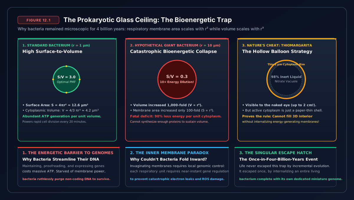
Why did prokaryotes never evolve true macroscopic complexity? In 2010, biochemist Nick Lane and evolutionary genomicist William Martin solved this fundamental enigma by calculating the bioenergetics of cell size:
In all prokaryotes, respiration is tied to the outer cell membrane. Protons are pumped across the plasma membrane, creating an electrochemical gradient that drives ATP synthesis. If a bacterium increases its radius by ten times, its internal volume (and energetic demand) expands by a factor of one thousand (V ∝ r^3), while its surface membrane area (and energy supply) expands by only a factor of one hundred (A ∝ r^2).
To overcome this surface-area deficit, a giant bacterium (such as Thiomargarita namibiensis) must fold its internal membranes and replicate thousands of copies of its entire genome along the periphery to locally monitor and regulate electron transport. A single giant bacterium may carry up to 20,000 copies of its full DNA genome! Nearly all of its energy budget is consumed simply replicating, transcribing, and translating identical copies of house-keeping genes. It has zero metabolic surplus left to evolve complex gene regulation, intracellular transport, or novel gene families.
The Endosymbiotic Revolution: The Merger of Two Worlds
In 1967, evolutionary biologist Lynn Margulis published her revolutionary paper “On the Origin of Mitosing Cells”, reviving the radical hypothesis of Serial Endosymbiosis. Margulis argued that the key internal powerhouses of eukaryotic cells—mitochondria and chloroplasts—were not built gradually from within, but were once free-living, independent prokaryotes engulfed by an ancient host cell.
For decades, Margulis’s theory was met with fierce academic skepticism. Today, genomic sequencing has transformed it into uncontested scientific fact:
- Mitochondria carry their own circular, bacterial DNA (mtDNA) and replicate independently inside our cells via binary fission.
- Mitochondrial ribosomes are bacterial-sized (70S) rather than eukaryotic (80S), and are sensitive to bacterial antibiotics (such as chloramphenicol and streptomycin).
- Mitochondria possess a double membrane, with the inner membrane containing cardiolipin—a signature lipid found only in bacterial membranes.
The Partners in the Merger: Asgard Archaea and Alphaproteobacteria
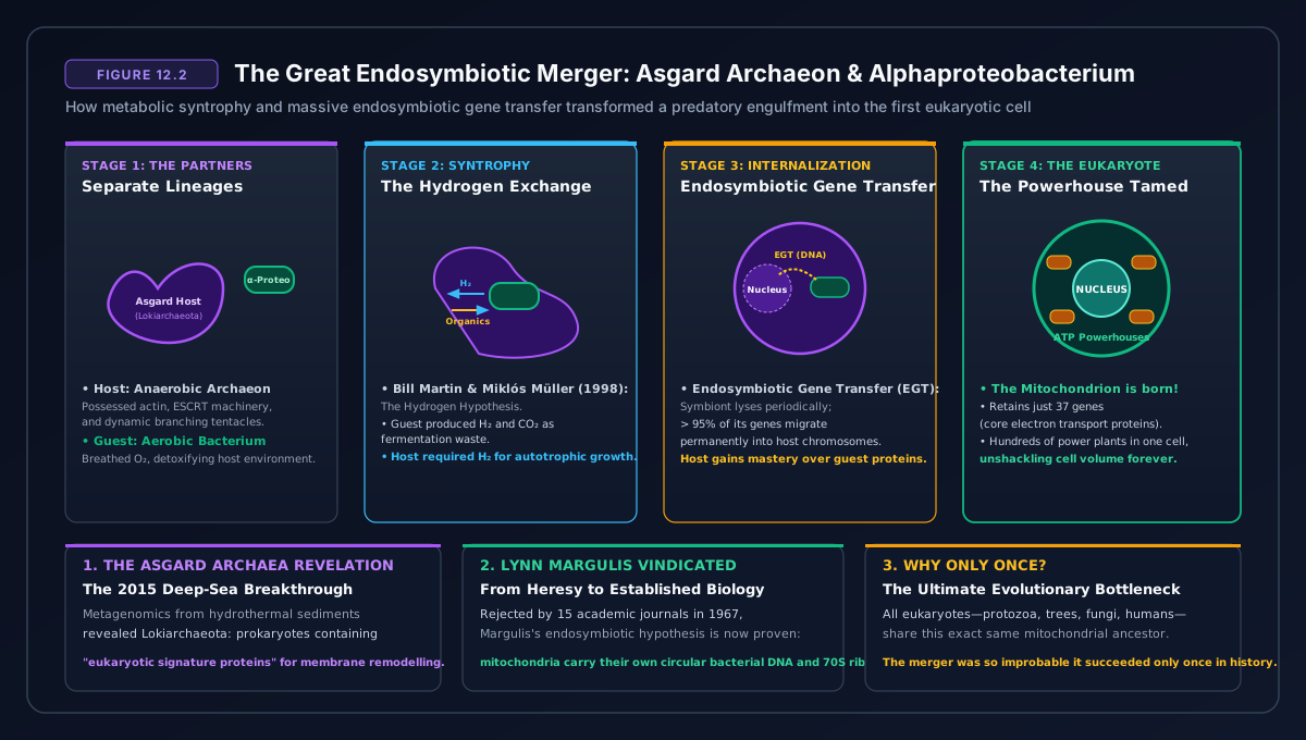
In 2015, metagenomic sequencing of hydrothermal ocean sediments near “Loki’s Castle” along the Arctic Mid-Atlantic Ridge identified a remarkable group of organisms: Asgard Archaea (Lokiarchaeota) (Spang et al. 2015). Genomic analysis proved that Asgard archaea are the closest living single-celled relatives of complex eukaryotic life: they encode eukaryotic signature proteins (ESPs) for actin-based cytoskeletons, membrane remodeling, and vesicle trafficking.
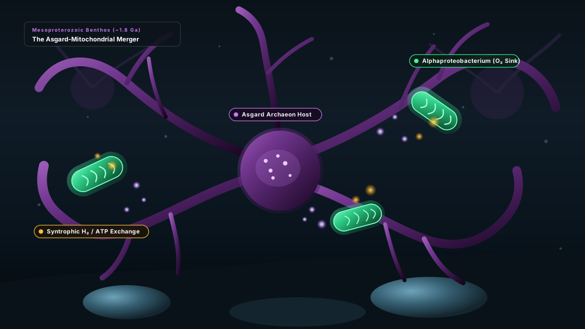
Roughly 1.8 billion years ago, an anaerobic Asgard-like archaeon formed a syntrophic metabolic partnership with an aerobic Alphaproteobacterium. The archaeon provided the bacterium with organic metabolic substrates; the bacterium, capable of aerobic respiration, detoxified oxygen and provided high-efficiency ATP. Eventually, the bacterium moved inside the archaeon’s cytoplasm—initiating the most consequential cohabitation in biological history.
The Energetic Breakthrough: 200,000 Times More Energy Per Gene
Once inside, the proto-mitochondrion underwent radical evolutionary streamlining: it transferred over 99% of its genes directly to the host nucleus via lateral gene transfer, retaining only a minimal, specialized genetic core (human mtDNA encodes just 13 essential respiratory proteins).
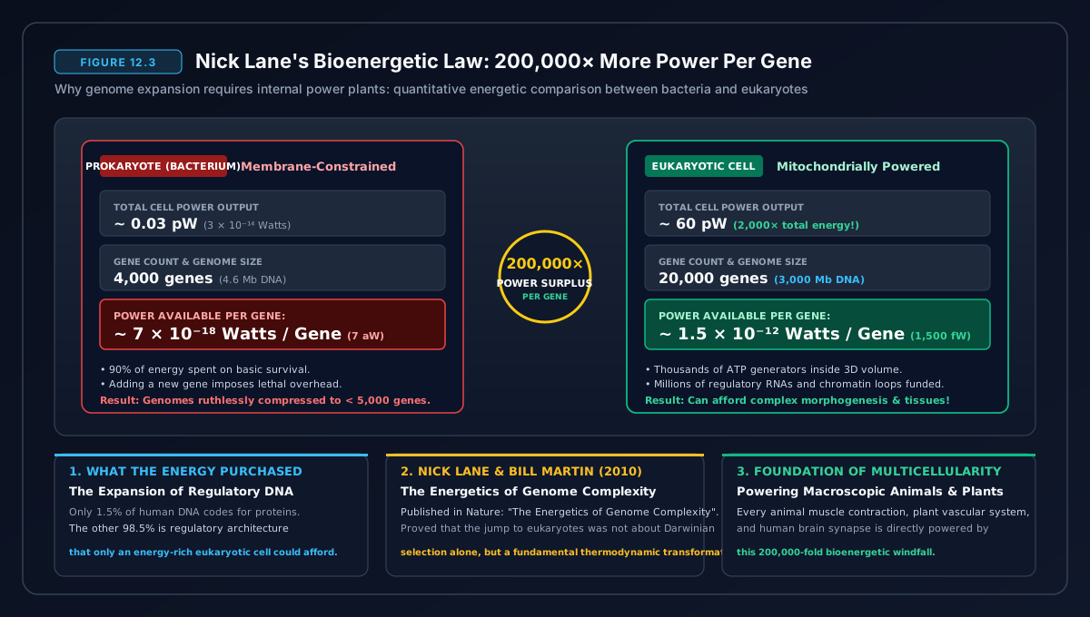
By delegating its genomic overhead to the central nucleus while packing hundreds or thousands of tiny, highly folded mitochondrial power plants into its cytoplasm, the eukaryotic cell shattered the surface-area-to-volume barrier forever:
$$\frac{\text{Power Available}}{\text{Gene}} \approx 200,000\times \text{ greater in Eukaryotes than in Prokaryotes}$$
With this staggering energy surplus, the eukaryotic cell could afford an enormous genome: thousands of novel regulatory genes, complex splicing machinery, signal transduction cascades, and internal motor proteins (kinesin and dynein) moving cargo along an intracellular highway of microtubules.
The Master Inventions: Nucleus, Introns, and the Cytoskeleton
The endosymbiotic merger triggered an explosion of cellular innovations that distinguish every eukaryote today:
1. The Nuclear Membrane & Spliceosomal Introns
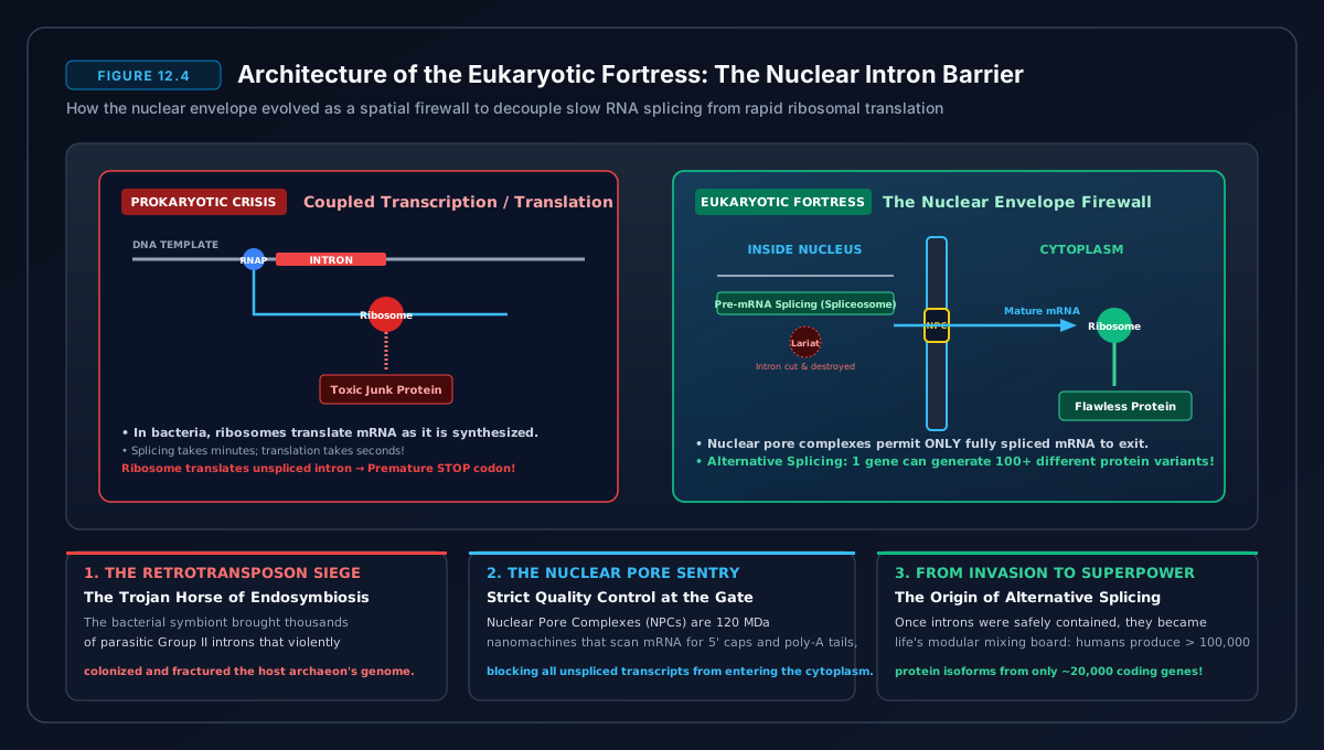
When thousands of bacterial endosymbionts lysed inside the host cytoplasm, their DNA fragmented, bombarding the host chromosomes with parasitic self-splicing Group II mobile introns. If an intron-carrying messenger RNA is translated immediately by ribosomes in the cytoplasm, it produces mangled, non-functional proteins.
The eukaryotic cell evolved the Nuclear Membrane as a spatial barrier: transcription and slow intron excision occur securely inside the nucleus via the spliceosome, and only completely verified, spliced mRNA transcripts are exported through nuclear pore complexes to ribosomes in the cytoplasm.
2. The Dynamic Cytoskeleton and Phagocytosis
Unlike rigid bacterial peptidoglycan cell walls, eukaryotes possess a flexible, dynamic internal scaffolding of actin microfilaments and tubulin microtubules. This allowed cells to change shape, engulf prey via phagocytosis, and establish directed intracellular vesicular transport.
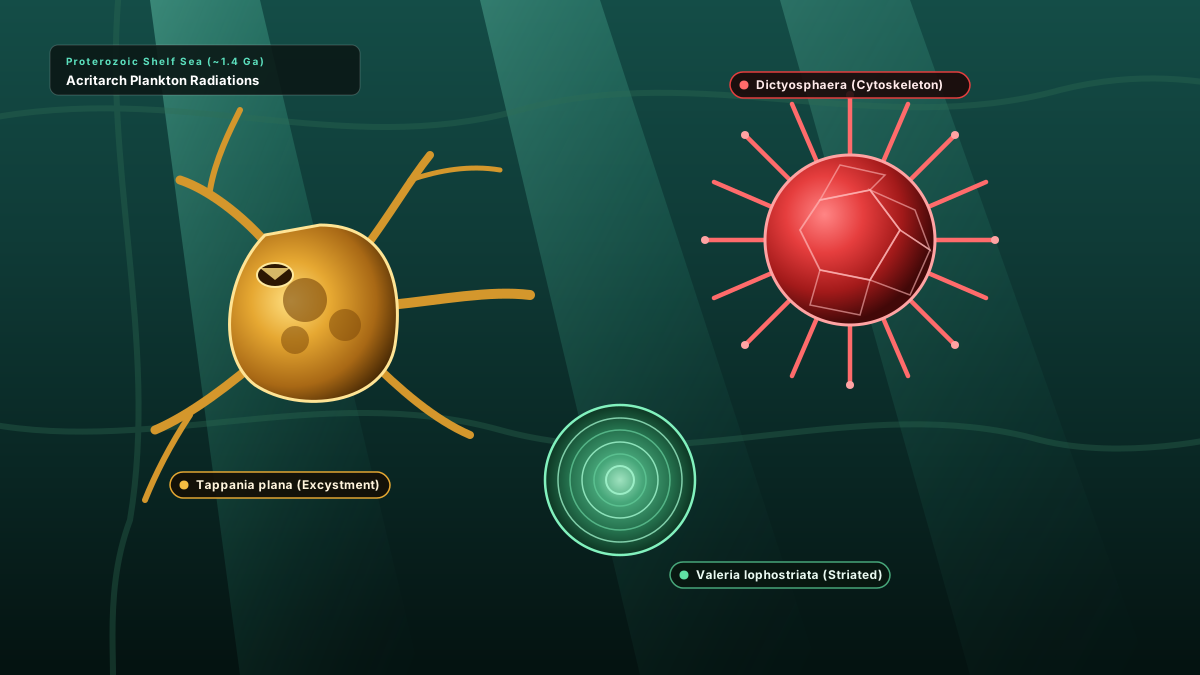
3. Secondary Endosymbiosis: The Green Revolution
Roughly 1.5 billion years ago, a eukaryotic cell containing mitochondria engulfed a free-living, photosynthetic Cyanobacterium. Rather than digesting it, the cell integrated it as an organelle: the Plastid (Chloroplast). This single evolutionary event birthed the entire Archaeplastida supergroup—the red algae, green algae, and the ancestors of all terrestrial plant life.
The Invention of Sex: Escaping Muller’s Ratchet
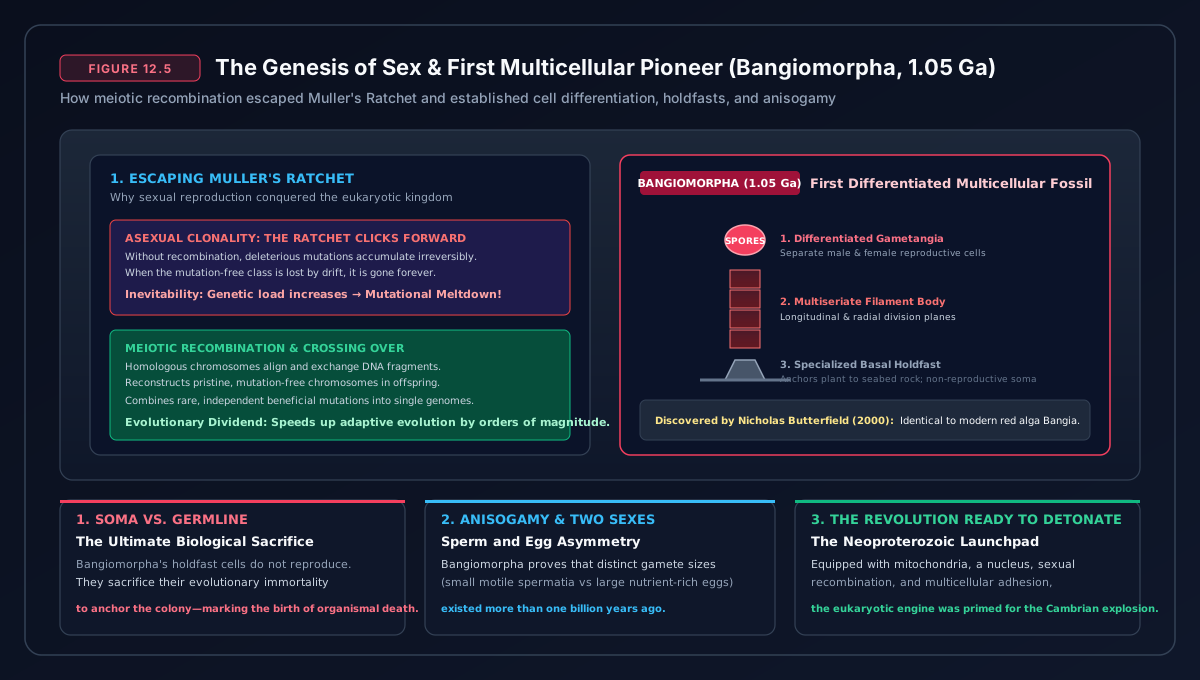
In asexual prokaryotic reproduction, a single cell divides into two identical clones. In sexual reproduction, two organisms combine half of their genetic material through Meiosis—a process involving homologous recombination and chromosomal crossing-over. At first glance, sex carries what evolutionary biologist John Maynard Smith called the “Twofold Cost of Sex”: a sexual female passes on only 50% of her genes to any individual offspring, and half of her offspring (males) cannot bear children directly.
Why did sex evolve and become the near-universal reproductive strategy of all complex eukaryotes? The answer lies in population genetics:
- Muller’s Ratchet: In an asexual population, deleterious genetic mutations accumulate irreversibly like clicks in a mechanical ratchet. Eventually, mutational load causes the lineage to decline into extinction. Sexual recombination allows genomes to shuffle, purging deleterious mutations and reconstructing mutation-free chromosomes.
- The Red Queen Hypothesis (Van Valen 1973): “It takes all the running you can do, to keep in the same place.” Parasites, viruses, and pathogens evolve thousands of times faster than multicellular hosts. Asexual clones present an identical, unchanging immunological target. Sexual recombination shuffles the host’s immune receptor alleles every single generation, preventing pathogens from wiping out an entire population.
The First Multicellular Pioneers: *Bangiomorpha pubescens*
For hundreds of millions of years during the mid-Proterozoic (often called the “Boring Billion” due to slow geochemical shifts, 1.8 to 0.8 Ga), single-celled eukaryotes diversified in the oceans. The earliest taxonomically confirmed macroscopic eukaryotic fossil is Bangiomorpha pubescens, discovered in 1.05-billion-year-old rocks on Somerset Island in arctic Canada (Butterfield 2000).
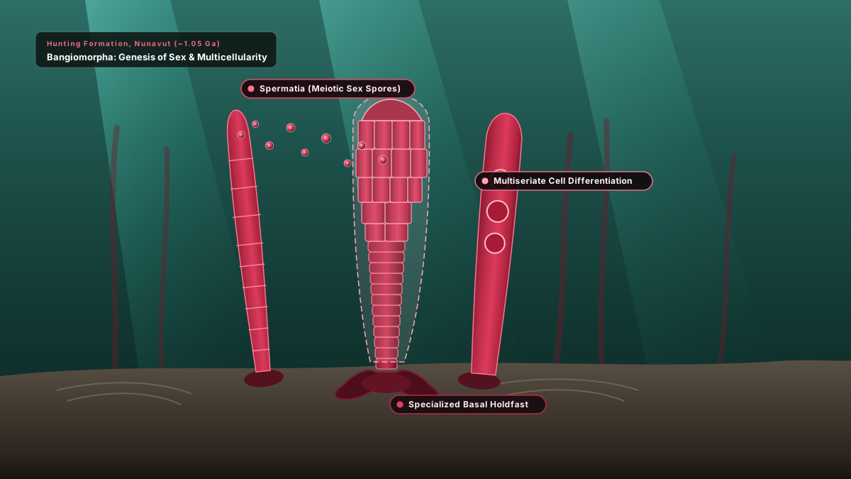
Bangiomorpha is a multicellular red alga that displays two world-changing biological innovations: cellular differentiation (possessing specialized discoid vegetative cells and an anchor holdfast cell) and true sexual reproduction (male and female spores). The genetic toolkit for building multicellular tissues—cell-to-cell adhesion (cadherins), intercellular communication (gap junctions), and developmental gene regulation—was officially assembled.
Prokaryotes vs. Eukaryotes: The Great Divide
| Biological Dimension | Prokaryotes (Bacteria & Archaea) | Eukaryotes (Animals, Plants, Fungi, Protists) |
|---|---|---|
| Energy Transduction | Across outer plasma membrane; surface-area limited | Across internal cristae of hundreds of mitochondria |
| Power per Gene | Baseline (1×) | 200,000× higher metabolic surplus |
| Genome Architecture | Single circular DNA; gene-dense; few non-coding introns | Multiple linear chromosomes; telomeres; extensive spliceosomal introns |
| Compartmentalization | No nucleus; simultaneous transcription/translation | Membrane-bound nucleus, endoplasmic reticulum, Golgi apparatus |
| Cytoskeleton & Motility | Rigid cell walls; external flagella rotary motors | Dynamic actin/tubulin cytoskeleton; phagocytosis; internal motor proteins |
| Reproductive Strategy | Asexual binary fission; lateral gene transfer | Meiotic sexual reproduction; homologous recombination |
| Morphological Limits | Strictly microscopic single cells or simple filaments | Complex macroscopic tissues, organ systems, and animals |
The Calm Before the Storm: The Neoproterozoic Threshold
By approximately 800 million years ago, the eukaryotic stage was set: single-celled and simple colonial eukaryotes possessed mitochondria, sexual recombination, complex genomes, and the intracellular machinery for tissue differentiation. Yet, large, macroscopic animals were still nowhere to be seen in the fossil record. Why?
The limiting factor was once again geochemical: atmospheric oxygen levels remained stalled at roughly 1% to 5% of present levels—sufficient for single cells, but far too low to support the deep oxygen diffusion required by thick, layered animal tissues. To smash open the gates of animal evolution, Earth required one final, catastrophic planetary convulsion.
In Part 13 of The Universal Chronicle, the planet will freeze into its deepest slumber and awaken in a riot of biological innovation: The Cambrian Crucible — Snowball Earth Thaw, Multicellular Body Plans, and the Apex Predators.
Academic Citations & Foundational Literature
- Sagan, L. (Margulis) (1967). “On the origin of mitosing cells.” Journal of Theoretical Biology, 14(3), 225–274. doi:10.1016/0022-5193(67)90079-3.
- Lane, N., & Martin, W. (2010). “The energetics of genome complexity.” Nature, 467, 929–936. doi:10.1038/nature09486.
- Spang, A., et al. (2015). “Complex archaea that bridge the gap between prokaryotes and eukaryotes.” Nature, 521, 173–179. doi:10.1038/nature14447.
- Butterfield, N. J. (2000). “Bangiomorpha pubescens n. gen., n. sp.: implications for the evolution of sex, multicellularity, and the Mesoproterozoic/Neoproterozoic radiation of eukaryotes.” Paleobiology, 26(3), 386–404. <a href="https://doi.org/10.1666/0094-8373(2000)0262.0.CO;2″ target=”_blank” rel=”noopener”>doi:10.1666/0094-8373(2000)0262.0.CO;2.
- Maynard Smith, J. (1978). The Evolution of Sex. Cambridge University Press. ISBN: 978-0521293020.
- Van Valen, L. (1973). “A new evolutionary law.” Evolutionary Theory, 1, 1–30.
- Koonin, E. V. (2010). “The origin and early evolution of eukaryotes in the light of phylogenomics.” Genome Biology, 11(5), 209. doi:10.1186/gb-2010-11-5-209.