Terrestrial bipedal locomotion is biologically extraordinary. Among all living mammals, only modern humans and our extinct hominin ancestors possess obligate, habitual striding bipedalism. Transforming an orthograde primate from a quadrupedal tree-dweller into an upright runner required a comprehensive mechanical overhaul of the axial skeleton, pelvis, lower limbs, and feet—a dynamic balancing act that trades biomechanical stability for unprecedented locomotor efficiency.
1. The Physics of the Striding Gait: The Inverted Pendulum Model
Human walking is modeled as an inverted pendulum. During the single-support phase of the gait cycle, the body’s center of mass (COM) arches upward over a stiff stance leg, converting kinetic energy ($E_k = frac{1}{2}mv^2$) into gravitational potential energy ($E_p = mgh$). As the body falls forward and downward into the double-support phase, potential energy is converted back into kinetic energy:
$$Delta E_{text{mech}} = |Delta E_k + Delta E_p|$$
In optimal bipedal walking, up to $70%$ of mechanical energy is recovered via this continuous conservative exchange, drastically reducing the net metabolic cost of transport ($C_{text{met}}$). In contrast, chimpanzee quadrupedalism and facultative bent-knee, bent-hip (BKBH) walking dissipate enormous energetic reserves through continuous eccentric muscular contraction.
2. The Axial Skeleton: S-Shaped Curvature & Center of Mass Alignment
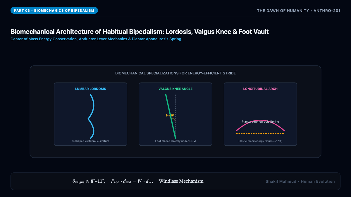
In quadrupedal primates, the vertebral column forms a single, continuous primary C-shaped thoracic arch. To balance an upright torso over the femoral heads without muscular fatigue, hominins evolved secondary spinal curvatures:
- Cervical Lordosis: Anterior convexity in the neck orienting the visual field forward horizontally.
- Thoracic Kyphosis: Posterior convexity accommodating thoracic organs and ribcage volume.
- Lumbar Lordosis: Severe anterior curvature of the lower spine (vertebrae L1–L5) that positions the torso’s center of gravity directly above the sacral promontory and acetabula (hip sockets). Wedging of the lower lumbar vertebrae ($L_4text{ and }L_5$) and intervertebral discs maintains this lordotic angle against vertical gravitational shear forces.
3. Pelvic Architecture & The Abductor Balancing Mechanism
The transformation of the hominin pelvis (the os coxae) represents the single greatest skeletal modification in human evolution. In non-human apes, the ilium is long, flat, and oriented in the coronal plane, extending upward along the lumbar spine. In hominins, the ilium underwent dramatic shortening and flaring into the sagittal plane:
This reorientation fundamentally altered the mechanical action of the lesser gluteal muscles (gluteus medius and gluteus minimus). In chimpanzees, these muscles function as hip extensors (propelling the body forward). In humans, their sagittal migration positions them as abductors. During the single-limb stance phase of walking, when the opposite leg swings forward, gravity exerts a torque tending to tilt the pelvis downward toward the unsupported side:
$$tau_{text{gravity}} = W_{text{body}} times d_W$$
The gluteus medius and minimus on the weight-bearing side immediately contract, generating an opposing counter-torque:
$$tau_{text{abductor}} = F_{text{abductor}} times d_{text{abductor}}$$
Equilibrium requires $tau_{text{abductor}} = tau_{text{gravity}}$, preventing pelvic collapse (the Trendelenburg sign) and ensuring smooth forward propulsion.
4. The Knee: The Bicondylar (Valgus) Angle
In apes, the femur is positioned vertically, perpendicular to the knee joint ($ ext{angle} approx 0^circ$). Because the hip joints are spaced widely apart, an ape walking bipedally must sway its entire upper body laterally from side to side to keep its center of mass over the weight-bearing foot. Hominins solved this problem through the evolution of the bicondylar (valgus) angle:
$$theta_{text{valgus}} approx 8^circtext{–}11^circ$$
The femoral shaft angles medially from the hip toward the midline of the body, placing the knees and feet directly beneath the body’s center of mass during stance. The lateral femoral condyle features an enlarged anterior lip (the patellar groove) that prevents lateral dislocation of the patella under the oblique quadriceps vector ($Qtext{-angle}$).
5. The Foot as an Elastic Energy Spring: Arches & The Windlass Mechanism
The human foot has surrendered all prehensile grasping capability to function as a rigid lever and elastic spring during the terminal stance and toe-off phases:
- Adducted Hallux: The great toe is brought permanently into parallel alignment with the other digits, losing its opposable joint mobility.
- Medial Longitudinal Arch: Supported by the calcaneus, talus, navicular, cuneiforms, and medial metatarsals.
- The Windlass Mechanism: The plantar aponeurosis is a thick band of connective tissue running from the calcaneus to the phalanges. During dorsiflexion of the toes prior to push-off ($> 30^circ$), the plantar fascia wraps tightly around the metatarsal heads, shortening the distance between heel and ball, elevating the longitudinal arch, and locking the tarsal bones into an impenetrable rigid lever that propels the body forward. During running, the elastic recoil of the Achilles tendon and plantar arch stores and returns up to $50%$ of mechanical energy per stride.